2024 SDMS Annual Conference Session Details
Morning Yoga | 0.75 SDMS CME Credit | Category: OT | Content Level: Beginner
Start your day with a 45-minute class that combines didactic information and movement. You'll learn stretching techniques to implement throughout your busy workday, helping to reduce stress and prevent musculoskeletal (MSK) injuries. This class is suitable for all levels, and mats will be provided. Comfortable clothing is recommended.
Objectives:
- Discuss the anatomy of the shoulder and arm.
- Understand the benefits of myofascial release and effective myofascial release practices for the shoulder and forearm.
- Learn stretching techniques to apply throughout the day to lessen tension and improve strength and mobility for the Sonographer.
Space is limited. Spots are available on a first come, first served basis.

Brandy Sundberg, MHPTT, RT (R), RDMS
Assistant Professor and Women’s Health Education Leader
University of Nebraska Medical Center and GE HealthCare
Brandy Sundberg is an Assistant Professor for the University of Nebraska Medical Center and Women's Health Education Leader for GE Healthcare. She has 20 years’ experience in Maternal Fetal Medicine where she was the Lead Sonographer, Sonographer Educator and Ultrasound Practitioner. Brandy graduated from the Diagnostic Medical Sonography program at the University of Nebraska Medical Center in 2002 with a Bachelor of Science in Radiation Science. She holds a Master’s Degree in Health Professions Teaching and Technology from the University of Nebraska Medical Center. Brandy is a Yoga Medicine trained yoga teacher and mindfulness meditation teacher. Brandy works with elite military professionals, healthcare professionals and individuals in her community to navigate stress and reduce burnout. Brandy has contributed to The Essential Guide to Yoga, focusing on the benefits of a restorative yoga practice. She is also an invited lecturer, discussing the benefits of mindfulness and stress reduction techniques for healthcare professionals, with a specific focus on alleviating musculoskeletal (MSK) injuries and burnout for sonographers. Brandy is co-founder of calmstrong.org, a website that provides functional movement and stress relief- created for healthcare professionals to integrate wellness into even the busiest of days.
Abdominal + Track | 1.0 SDMS CME Credit | Category: OT | Content Level: Beginner
Sonography, a field vital to patient care, faces the risk of losing passionate professionals to burnout. This presentation explores the delicate balance between the technical demands of the profession and the emotional connection sonographers seek in their work. Attendees will gain insights into fostering a workplace environment that not only addresses burnout causes, but also actively nurtures the passion that drew sonographers to the profession in the first place.
Objectives:
- Identify at least three strategies for recognizing and sustaining the passion of sonographers in their daily work.
- Explain the importance of integrating, empathy, and compassion into the technical aspects of sonography to enhance patient care.
- Outline a preliminary action plan for creating a passion-driven culture within their workplace.

Jennifer Lindsey
Advanced Imaging Inc.
Co-Founder
Jennifer Lindsey is the Co-Founder and Lead Business Advisor at Advanced Imaging, Inc. She and her business partner launched their ultrasound facility over 20 years ago, and since then have scaled their business to include multiple diagnostic ultrasound service options, business coaching and consulting in the sonography field, ultrasound equipment, and ultrasound education services. Fueled by a deep-seated passion for the pivotal role sonography plays in healthcare, Jennifer is unwavering in her dedication to mentoring sonographers, guiding them toward achieving excellence in their careers—whether they aspire to excel as employees or to thrive as practitioners running their own businesses.
Cardiac Track | 1.0 SDMS CME Credit | Category: AE | Content Level: Intermediate
This presentation will highlight approaches to best practices in education, QI, lab accreditation, and new technology for the echo lab.
Objectives:
- Provide information on opportunities for education in the echo lab.
- Describe the lab accreditation process and standards for clinical practice and quality improvements.
- Apply new technology that helps to improve quality and workflow in the echo lab.
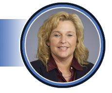
Pam Burgess, BS, ACS, RDCS, RDMS, RVT, FASE
Clinical Manager
Atrium Health Wake Forest Baptist
Pam has been a clinical sonographer for many years and a speaker and course director for several cardiac ultrasound courses. She is the immediate past president of IAC Echo division and serves on the All IAC BOD. She has also served on the ASE BOD, various committees, and past ASE program chair.
OB/GYN Track | 1.0 SDMS CME Credit | Category: FE | Content Level: Intermediate
This presentation will review the anatomy of a dilated coronary sinus and a primum ASD and showcase the associated findings for each defect to help determine the diagnosis.
Objectives:
- Identify a dilated coronary sinus and associated pathology.
- Identify a primum atrial septal defect and associated pathology.
- Review associated findings with a dilated coronary sinus and a primum ASD to help determine diagnosis.
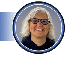
Mary Davis, RDCS (AE, PE, FE), RCS, FASE
Pediatric Fetal Echo Lab Supervisor
UC Davis Medical Center
Mary Davis is a seasoned pediatric/fetal cardiac sonographer with a career spanning over two decades. She began her journey on the east coast in 1999 and played a pivotal role as one of the founding members of the South Florida Echo Society in Palm Beach County during the early 2000s. Driven by her passion for congenital cardiac imaging, Mary relocated to Phoenix, Arizona, where she joined Phoenix Children's Hospital. Over the course of 13 years, she dedicated herself to the program, assuming leadership roles and eventually becoming the manager of Phoenix Children's CardioDiagnostics. In this role, Mary oversaw three departments and managed approximately 50 employees. Continuing her commitment to education and advancement in the field, Mary has been a valued faculty member of the Phoenix Fetal Cardiology Symposium every year. In 2021, seeking a better work/life balance, Mary made the decision to relocate to Sacramento, California, where she currently serves as a Sonographer at UC Davis Medical Center. Her wealth of experience and dedication to pediatric and fetal cardiac sonography continue to make a significant impact in the field.
Vascular Track | 1.0 SDMS CME Credit | Category: VT | Content Level: Intermediate
This presentation will describe unusual findings encountered in patients presenting for sonographic assessment of the extracranial carotid arteries. Atherosclerotic and non-atherosclerotic pathologies will be discussed. Cases will include carotid dissection, aneurysms, carotid webs, unanticipated flow dynamics and traumatic injuries.
Objectives:
- Identify a range of uncommon atherosclerotic and non-atherosclerotic pathologies that can present in the carotid arteries.
- Recognize the sonographic features of each pathology.
- Interpret the sonographic and pathophysiological findings to present accurate diagnostic information.
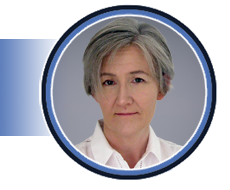
Jacqui Robinson, RN, RVT, AMS, FASA
Chief Vascular Sonographer
Liverpool Hospital
Jacqui Robinson serves as the Chief Vascular Sonographer at Liverpool Hospital, a prominent teaching hospital in South-West Sydney, Australia. With a career in vascular ultrasound spanning back to 1989, Jacqui has amassed extensive experience in sonography education, clinical ultrasound practice, and cardiovascular research. Throughout her career, Jacqui has held numerous executive committee positions with the Australasian Society for Ultrasound in Medicine and the Australasian Sonographers Association. She is actively involved in teaching and clinically supervising students from accredited graduate sonography programs across Australia, with a particular interest in rural and remote education. Jacqui is a frequent presenter at local, national, and international conferences, contributing her expertise to the advancement of the field. In recognition of her professional excellence and dedicated service to the sonography profession, Jacqui was honored with a Fellowship from the Australasian Sonographers Association in 2019.
Abdominal + Track | 1.0 SDMS CME Credit | Category: AB | Content Level: Intermediate
Contrast Enhanced Ultrasound (CEUS) is gaining momentum as a unique diagnostic specialty in the field of abdominal ultrasound. The focus of this presentation will be on the role of microbubble contrast agents in liver imaging today. Special attention will be given to contrast agent applications, image acquisition, software requirement, and how to get the images.
Objectives:
- Review Microbubble contrast agents, and their application in liver imaging.
- Review techniques and current imaging protocol, including the difference between contrast agents in ultrasound vs. CT and MRI.
- Identify why the liver is a unique organ amenable to the diagnostic use of ultrasound contrast agents.
- Understand Image Acquisition techniques and see how microbubble contrast agents affect how we image.

Christina Merrill, BSc, RDMS, CRGS, CRVS
Sonographer
Alberta Health Services
Christina Merrill, BSc, RDMS, CRGS, CRVS, serves as the Lead Sonographer for Dr. Stephanie Wilson at Foothills Medical Center in Alberta, Canada. With 15 years of specialization in Contrast Enhanced Ultrasound (CEUS), she is a member of the board of directors for the International Contrast Ultrasound Society (ICUS), where she teaches CEUS globally, emphasizing safe contrast agent utilization. Christina actively engages in clinical research, publishing and collaborating on investigations involving microbubble contrast agents for medical imaging, particularly in diagnosing and characterizing liver tumors.
Cardiac Track | 1.0 SDMS CME Credit | Category: AE | Content Level: Intermediate
This lecture will review the basics of EKGs and rhythms and how they affect the heart and disease process.
Objectives:
- Review basic EKGs and rhythms for the cardiac sonographer.
- Develop an understanding of how different EKG findings can help you make the diagnosis of different heart diseases.
- Look at common associated EKG changes related to different pathologies.

Richard Palma, BS, ACS, RCCS, RCS, FACVP, FSDMS, FASE
Director and Clinical Coordinator
Duke University School of Medicine
Richard (Richie) Palma, BS, ACS, RCS, RDCS, FACVP, FSDMS, FASE, is the founder, director, and clinical coordinator of Duke’s Cardiac Ultrasound Certificate Program. He received the 2021 Duke School of Medicine Cardiology Teaching Award and is the only man to have received both the "Distinguished Teaching Award" from the SDMS and ASE. Appointed to the CAAHEP Board of Directors in spring 2022, Richie also serves on the Board of Directors for the SDMS and the SDMS Foundation (2021-2025). He contributes extensively to the sonography profession as an educator, author, program developer, and national and international speaker on sonography and echocardiography. Richie authored the fifth edition of Echocardiographer’s Pocket Reference, a definitive reference since 1993. He succeeded Sid Edelman at ESP Ultrasound and is a national speaker for Philips and Lantheus Medical Imaging. A Fellow of the SDMS, the American Society of Echocardiography, and the Alliance of Cardiovascular Professionals, Richie achieved ACS certification in 1990, marking the beginning of his sonography career.
OB/GYN Track | 1.0 SDMS CME Credit | Category: OB | Content Level: Beginner
The purpose of this presentation is to familiarize the sonographer with the cavum septi pellucidi (CSP), review the anatomy and ultrasound image acquisition, and stress the clinical importance of documenting the presence of this structure during the routine anatomic survey of the fetus. The congenital absence of the CSP will be reviewed as well as recommendations for the management and follow-up of this condition.
Objectives:
- Recognize the anatomic location and normal sonographic appearance of the cavum septi pellucidi.
- Describe potential anomalies that may be associated with absence of the cavum septi pellucidi.
- Discuss options for reporting abnormal findings and promoting appropriate follow-up in cases of congenital absence of the CSP.

Lisa M. Allen, BS, RDMS, RDCS, RVT, FAIUM
Ultrasound Coordinator / Clinical Instructor / Clinical Sonographer
SUNY Upstate Medical University
Lisa M. Allen has been a clinical sonographer and educator at the Regional Perinatal Center at the SUNY Upstate Medical University since 1992. She earned her Bachelor of Science degree from Rochester Institute of Technology where, in 2018, she received a Distinguished Alumna Award through the College of Health Sciences and Technology. Lisa has worked with the Society of Diagnostic Medical Sonography on the Continuing Medical Education Review Committee, the Advanced Practice Committee, and the National Certification Exam Review Task Force. She has served on numerous committees for the American Institute of Ultrasound in Medicine and has served two terms as the Second Vice President for this organization. In 2015, Lisa received the AIUM Distinguished Sonographer Award. She has served the American Registry for Diagnostic Medical Sonography on the Obstetrics and Gynecology Exam Development Task Force and the Recertification Oversight Task Force Committee, was a subject matter in fetal echocardiography and obstetrics and gynecology. Lisa has numerous publications on the prenatal diagnosis of congenital fetal anomalies.
Vascular Track | 1.0 SDMS CME Credit | Category: VT | Content Level: Intermediate
Imaging plays a crucial role in the evaluation and management of acute deep venous thrombosis (DVT) and pulmonary embolism (PE). This presentation will explore current methods utilized for the diagnosis and treatment of DVT and PE.
Objectives:
- Review risk factors associated with deep venous thrombosis and pulmonary embolism.
- Analyze ultrasound and CT imaging techniques utilized for diagnosis of deep venous thrombosis and pulmonary embolism.
- Discuss treatment methods including medical management, catheter-directed thrombolysis, and percutaneous suction thrombectomy.
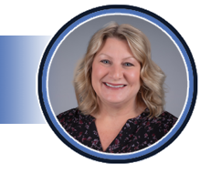
Amy Wilson, MS, RT(R), RDMS (AB, OB, BR), RVT
Chair and Clinical Professor, Diagnostic Medical Sonography
University of Southern Indiana
Amy Wilson serves as the Chair and Clinical Professor of the Diagnostic Medical Sonography program at the University of Southern Indiana in Evansville, Indiana. With over 25 years of experience in sonography, Amy has been a member of the Society of Diagnostic Medical Sonography (SDMS) since 2005. Amy holds a Master of Science in Education degree from the University of Southern Indiana, a Bachelor of Science in Medical Imaging Technology degree, and an Associate of Science in Radiologic Technology degree from Indiana University School of Medicine. Throughout her career, Amy has been actively involved in the SDMS, contributing to committees such as the Government Relations Committee and the Education Committee. Amy has also held positions as Director on the SDMS Board of Directors and SDMS Foundation Board of Directors. She served as the board liaison to the SDMS Foundation Emerging Leaders Program from 2020 to 2022. Currently, Amy serves as the Secretary for both the SDMS and SDMS Foundation. In addition to her administrative roles, Amy has contributed significantly to education and research in the field of diagnostic medical sonography. She has created over 20 SDMS webinars, presented at the SDMS Annual Conference, and authored several articles published in the Journal of Diagnostic Medical Sonography (JDMS). In recognition of her outstanding research and review articles, Amy received the second place Kenneth R. Gottesfeld Award in 2011.
Abdominal + Track | 1.0 SDMS CME Credit | Category: OT | Content Level: Beginner
As the demands on sonographers intensify in today's dynamic healthcare setting, the toll on their well-being becomes a pressing concern. This session delves into the nuanced challenges sonographers face, exploring not only the root causes of burnout but also the psychological impact on their overall health. Practices and sonographers alike will gain insights into fostering a resilient, positive workplace culture, ensuring that the human element of sonography doesn't get lost in the diagnostic process.
Objectives:
- List three aspects contributing to sonographer burnout.
- Explain the psychological impact of burnout on the overall health of sonographers.
- Outline a personalized plan incorporating at least three emotional well-being strategies to maintain mental health and emotional resilience in their own demanding roles as sonographers.

Jennifer Lindsey
Advanced Imaging Inc.
Co-Founder
Jennifer Lindsey is the Co-Founder and Lead Business Advisor at Advanced Imaging, Inc. She and her business partner launched their ultrasound facility over 20 years ago, and since then have scaled their business to include multiple diagnostic ultrasound service options, business coaching and consulting in the sonography field, ultrasound equipment, and ultrasound education services. Fueled by a deep-seated passion for the pivotal role sonography plays in healthcare, Jennifer is unwavering in her dedication to mentoring sonographers, guiding them toward achieving excellence in their careers—whether they aspire to excel as employees or to thrive as practitioners running their own businesses.
Cardiac Track | 1.0 SDMS CME Credit | Category: AE | Content Level: Advanced
This presentation will provide an overview of echo assessment of valvular disease and the hemodynamic impact when mixed valve disease is present.
Objectives:
- Define the role of echo assessment of valvular heart disease.
- Discuss the impact on hemodynamic echo assessment of mixed valvular disease.
- Provide case examples of echocardiographic assessment of mixed valve disease.

Pam Burgess, BS, ACS, RDCS, RDMS, RVT, FASE
Clinical Manager
Atrium Health Wake Forest Baptist
Pam has been a clinical sonographer for many years and a speaker and course director for several cardiac ultrasound courses. She is the immediate past president of IAC Echo division and serves on the All IAC BOD. She has also served on the ASE BOD, various committees, and past ASE program chair.
OB/GYN Track | 1.0 SDMS CME Credit | Category: FE | Content Level: Intermediate
This presentation will review the anatomy and physiology of Tetralogy of Fallot variants in fetal echo.
Objectives:
- Review the embryologic development of a Tetralogy variant.
- Understand the differences across the Tetralogy variants.
- Review the Tetralogy variants and which patients require immediate imaging to determine if medication is needed to keep the duct open.

Mary Davis, RDCS (AE, PE, FE), RCS, FASE
Pediatric Fetal Echo Lab Supervisor
UC Davis Medical Center
Mary Davis is a seasoned pediatric/fetal cardiac sonographer with a career spanning over two decades. She began her journey on the east coast in 1999 and played a pivotal role as one of the founding members of the South Florida Echo Society in Palm Beach County during the early 2000s. Driven by her passion for congenital cardiac imaging, Mary relocated to Phoenix, Arizona, where she joined Phoenix Children's Hospital. Over the course of 13 years, she dedicated herself to the program, assuming leadership roles and eventually becoming the manager of Phoenix Children's CardioDiagnostics. In this role, Mary oversaw three departments and managed approximately 50 employees. Continuing her commitment to education and advancement in the field, Mary has been a valued faculty member of the Phoenix Fetal Cardiology Symposium every year. In 2021, seeking a better work/life balance, Mary made the decision to relocate to Sacramento, California, where she currently serves as a Sonographer at UC Davis Medical Center. Her wealth of experience and dedication to pediatric and fetal cardiac sonography continue to make a significant impact in the field.
Vascular Track | 1.0 SDMS CME Credit | Category: VT | Content Level: Intermediate
A functional access is the dialysis-dependent patients’ lifeline, with many undergoing multiple interventions or revisions over time. This presentation will take a “Why, What, and How” approach to help build confidence in how sonographers might approach these often-challenging studies.
Objectives:
- Describe the anatomical and hemodynamic features of surgically created vascular access for hemodialysis.
- Review and interpret the indications for ultrasound imaging in hemodialysis access.
- Interpret the sonographic features of access dysfunction and present diagnostic information to support patient management.

Jacqui Robinson, RN, RVT, AMS, FASA
Chief Vascular Sonographer
Liverpool Hospital
Jacqui Robinson serves as the Chief Vascular Sonographer at Liverpool Hospital, a prominent teaching hospital in South-West Sydney, Australia. With a career in vascular ultrasound spanning back to 1989, Jacqui has amassed extensive experience in sonography education, clinical ultrasound practice, and cardiovascular research. Throughout her career, Jacqui has held numerous executive committee positions with the Australasian Society for Ultrasound in Medicine and the Australasian Sonographers Association. She is actively involved in teaching and clinically supervising students from accredited graduate sonography programs across Australia, with a particular interest in rural and remote education. Jacqui is a frequent presenter at local, national, and international conferences, contributing her expertise to the advancement of the field. In recognition of her professional excellence and dedicated service to the sonography profession, Jacqui was honored with a Fellowship from the Australasian Sonographers Association in 2019.
Abdominal + Track | 1.0 SDMS CME Credit | Category: AB | Content Level: Intermediate
Focal liver lesions are often encountered on greyscale imaging and their noninvasive diagnosis is necessary, especially if they are atypical. Imaging characterization of focal liver lesions and exclusion of malignancy are of prime importance. This presentation will review an established algorithm for diagnosis of liver masses on CEUS. It will include a step-by-step approach for the diagnosis of focal liver lesions with CEUS. For the first time, Contrast Enhanced Ultrasound becomes competitive with CT and MRI!
Objectives:
- Review the properties of contrast agents within the liver including liver phases and their importance in imaging.
- Understand the importance of specific enhancement patterns in liver tumors including the importance of washout and provide a pictorial review of the CEUS algorithm for diagnosis.
- Apply the learned algorithm in a fun test your knowledge section.

Christina Merrill, BSc, RDMS, CRGS, CRVS
Sonographer
Alberta Health Services
Christina Merrill, BSc, RDMS, CRGS, CRVS, serves as the Lead Sonographer for Dr. Stephanie Wilson at Foothills Medical Center in Alberta, Canada. With 15 years of specialization in Contrast Enhanced Ultrasound (CEUS), she is a member of the board of directors for the International Contrast Ultrasound Society (ICUS), where she teaches CEUS globally, emphasizing safe contrast agent utilization. Christina actively engages in clinical research, publishing and collaborating on investigations involving microbubble contrast agents for medical imaging, particularly in diagnosing and characterizing liver tumors.
Cardiac Track | 1.0 SDMS CME Credit | Category: AE | Content Level: Beginner
When conducting an echocardiogram, the mental tools we bring to the procedure are the standard lab protocol, the diagnosis, and the patient's history. This presentation will address how these three elements contribute to our ability to perform the most effective echocardiogram for patients.
Objectives:
- Understand the importance of having a plan before beginning an echocardiogram.
- Recognize how a diagnosis and patient history provide insight into what to look for during the echocardiogram.
- Acquire tips on adjusting protocols based on time constraints or findings during the echocardiogram.

Jennifer Cruz, RDCS
Cardiac Sonographer Lead
Inova Fairfax Hospital
Jennifer Cruz is a seasoned professional in the realm of Cardiac Sonography, boasting an impressive 17-year tenure within diverse cardiology labs. As the lead at Inova Fairfax hospital in Virginia, Jennifer plays a pivotal role in enhancing the department’s efficiency and quality standards. With a keen focus on perfecting protocols and driving quality improvement initiatives, Jennifer ensures that fellow sonographers deliver exceptional patient care. However, Jennifer’s expertise extends beyond administrative duties. She possesses a profound passion for structural heart space. Whether it’s conducting scans, refining techniques, or educating others, she is deeply committed to advancing the field of cardiac sonography. Her vast experience serves as a foundation for guiding and mentoring fellow sonographers, empowering them to thrive in this specialized area.
OB/GYN Track | 1.0 SDMS CME Credit | Category: OB | Content Level: Advanced
Endometriosis is a disease that affects 10% or more of reproductive age women in the United States and is the leading cause of infertility. While patients may present with pain, they may also be asymptomatic and unaware of their disease. In fact, the average delay in diagnosis in the USA is 10 years! By implementing an Augmented exam with a few additional maneuvers (as suggested by the SRU Consensus), developing a bowel prepped exam for detailed disease mapping and MRI directed ultrasound exams for surgical planning we will transform patient care and help strength the future of families in our country.
Objectives:
- Provide Background on Endometriosis and the role of Radiology in the management of Endometriosis.
- Review of Multidisciplinary rounds at our institution.
- Review Dedicated US protocols, cases, and reporting (with MRI correlation).

Wendaline VanBuren, MD
Radiologist, Associate Professor of Radiology
Mayo Clinic
Dr. VanBuren is a board-certified radiologist, subspecialized in ultrasound, abdominopelvic and breast imaging. Dr. VanBuren is the chair of the Gynecological imaging section, with a dedicated interest in Endometriosis. Dr. VanBuren is one of the founding Directors for the Mayo Clinic Gynecological and Breast Imaging course. On a national level, she has co-founded and chaired the Disease Focused Panel on Endometriosis for the Society of Abdominal Radiology and was the invited guest editor for the journal Abdominal Radiology Special Edition on Endometriosis. Her ongoing work is frequently highlighted at a variety of national and international radiology and gynecology conferences. Dr. VanBuren takes pride in advocacy and propagating a passion for medical imaging, education, clinical and scientific collaboration.
Vascular Track | 1.0 SDMS CME Credit | Category: VT | Content Level: Intermediate
This presentation will enhance comprehension of Nutcracker Syndrome and Nutcracker Physiology. Attendees will review renal vein compression anatomy and its link to pelvic congestion. The presenter will offer a scanning method to better visualize the Gonadal vein.
Objectives:
- Review patient demographic and symptoms associated with Nutcrackers.
- Provide a suggested protocol and interpretation for reporting our lab has recently developed. Helpful hints on image optimization.
- Review case presentations on normal and abnormal study, post-operative studies, and treatment Options.
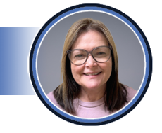
Tammy Rubin, RDMS, RVT
Lead Sonographer in the Vascular Lab for Vascular Surgery and Cardiovascular Medicine
Cleveland Clinic Heart Vascular and Thoracic Institute (HVTI)
Tammy Rubin is an experienced Vascular Sonographer with a thirty-year tenure at the Cleveland Clinic Heart, Vascular, and Thoracic Institute, specializing in vascular ultrasound for the past twenty-five years. For the last fifteen years, she has served as the Lead Sonographer in the Vascular Surgery Department, contributing to research, implementing new ultrasound protocols, updating structured reporting, and educating staff, students, residents, fellows, visiting physicians, and sonographers. Tammy provides expertise in pre- and post-operative patient revascularization complexity. She authored a book chapter on "Aorta and Iliac Arteries" for The Vascular System (Diagnostic Medical Sonography Series) Third Edition by Ann Marie Kupinski. A long-standing member of the Society of Diagnostic Medical Sonography (SDMS), Tammy has spoken at the SDMS Annual Conference and participated in refresher courses. She has excelled, winning first place in the Sonographer Poster Competition at the Annual Conference. Tammy's involvement with the Intersectional Accreditation Commission (IAC VL) as a Vein Center reviewer, vascular ultrasound reviewer, and webinar presenter has been influential. She has presented for various organizations in Ohio, crediting her vascular knowledge to supportive colleagues and physicians who facilitated her advancement to lead sonographer in vascular surgery.
Abdominal + Track | 1.0 SDMS CME Credit | Category: PS | Content Level: Intermediate
This lecture will provide an overview of the role of sonography in the evaluation of the pediatric neck with focus on the tonsils and vocal cords. Sonographic anatomy will be described, and anatomy and pathology will be illustrated. The goals of this exhibit are: 1. Describe the technical approach of performing pediatric neck sonography 2. Highlight common exam indications 3. Identify and describe the sonographic appearance of normal variants 4. Review common and rare lesions in pediatric patients 5. Illustrate the sonographic findings and their correlation with other modalities, as appropriate 6. Discuss diagnostic criteria of imaging findings.
Objectives:
- Understand the basics of tonsillar anatomy and pathology.
- Understand the basics of vocal cord anatomy and pathology.
- Describe the techniques and protocol for imaging vocal cords and tonsils using ultrasound.

Tara Cielma, BS, RT(S), RDMS, RDCS, RVT, FSDMS
Sonography Research Specialist
Children's National Hospital
With over 16 years of experience, Tara is a well-established leader in the sonography field. Her credentials include abdomen, OB/GYN, neurosonography, vascular technology, fetal echo, and a credential from ARRT in Sonography. She is also designated as an SDMS Fellow.
Tara's dedication to advancing the field of sonography extends beyond her professional roles; she is deeply committed to giving back to the sonography community. Tara actively volunteers with SDMS, ARDMS/Inteleos, and SPR (just to name a few). She also reviews manuscripts for the Journal of Diagnostic Medical Sonography (JDMS), contributes to writing registry items and preparation material, and provides hands-on workshops for facilities seeking to develop or enhance their pediatric and fetal imaging skillset.
Tara has numerous chapters, journal articles, and a co-authored pediatric registry review book to her name. Tara's extensive experience, unwavering dedication, and passion for volunteerism ensure she will be making waves in the field for years to come.
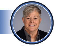
Monique Riemann, RDMS, RVT
Research Sonographer
Phoenix Children’s Hospital
Monique Riemann is an ultrasound technologist with 37 years of experience in the field. Her registries include Abdomen, OB/GYN, Vascular, (Neuro), and Pediatric. She began her career at Jackson Memorial Medical Center in Miami, Florida, before relocating to Arizona. After many years in a lead position at Phoenix Children’s Hospital, she transitioned to a research position where she has played a significant role in creating ultrasound exams that were once radiation-based, utilizing the motto "Think Ultrasound First." Monique has received awards for scientific presentations and poster competitions at the Society of Diagnostic Medical Sonography (SDMS). Over the last 7 years, she has had the pleasure of speaking at several conferences. To date, she has over 20 publications on various topics related to ultrasound. Monique's passion for ultrasound extends beyond her research role; she is also employed part-time as an educator and lab instructor at West Coast Ultrasound Institute, where she contributes to the development of future sonographers.
Cardiac Track | 1.0 SDMS CME Credit | Category: AE | Content Level: Beginner
With the cardiac field expanding due to structural heart procedures potentially leading to obstructions, and with an increasing number of patients diagnosed with HCM, distinguishing between an increased LV gradient and LVOT obstruction, or identifying cases where a patient may have both, has become increasingly crucial in echocardiograms. This presentation will review LVOT Obstruction and LV Gradient as well as cases with both.
Objectives:
- Identify the common reasons a patient will have an LVOT Obstruction, Increased LV Gradient, or both.
- Identify what increased LV gradient and LVOT obstruction look like.
- Provide tips on how to alter a protocol depending on time constraint or what is discovered while performing the echocardiogram.

Jennifer Cruz, RDCS
Cardiac Sonographer Lead
Inova Fairfax Hospital
Jennifer Cruz is a seasoned professional in the realm of Cardiac Sonography, boasting an impressive 17-year tenure within diverse cardiology labs. As the lead at Inova Fairfax hospital in Virginia, Jennifer plays a pivotal role in enhancing the department’s efficiency and quality standards. With a keen focus on perfecting protocols and driving quality improvement initiatives, Jennifer ensures that fellow sonographers deliver exceptional patient care. However, Jennifer’s expertise extends beyond administrative duties. She possesses a profound passion for structural heart space. Whether it’s conducting scans, refining techniques, or educating others, she is deeply committed to advancing the field of cardiac sonography. Her vast experience serves as a foundation for guiding and mentoring fellow sonographers, empowering them to thrive in this specialized area.
OB/GYN Track | 1.0 SDMS CME Credit | Category: OB | Content Level: Intermediate
This presentation aims to educate the sonographer about the prenatal diagnosis of hypospadias. It will review the embryology, incidence and types of hypospadias. In addition, it will briefly review anomalies of the external male genitalia that are closely associated with this condition.
Objectives:
- Define hypospadias and the various types of this congenital abnormality.
- Review the role of prenatal sonography in the diagnosis of this condition.
- Discuss the embryology, incidence and anomalies closely associated with hypospadias.

Lisa M. Allen, BS, RDMS, RDCS, RVT, FAIUM
Ultrasound Coordinator / Clinical Instructor / Clinical Sonographer
SUNY Upstate Medical University
Lisa M. Allen has been a clinical sonographer and educator at the Regional Perinatal Center at the SUNY Upstate Medical University since 1992. She earned her Bachelor of Science degree from Rochester Institute of Technology where, in 2018, she received a Distinguished Alumna Award through the College of Health Sciences and Technology. Lisa has worked with the Society of Diagnostic Medical Sonography on the Continuing Medical Education Review Committee, the Advanced Practice Committee, and the National Certification Exam Review Task Force. She has served on numerous committees for the American Institute of Ultrasound in Medicine and has served two terms as the Second Vice President for this organization. In 2015, Lisa received the AIUM Distinguished Sonographer Award. She has served the American Registry for Diagnostic Medical Sonography on the Obstetrics and Gynecology Exam Development Task Force and the Recertification Oversight Task Force Committee, was a subject matter in fetal echocardiography and obstetrics and gynecology. Lisa has numerous publications on the prenatal diagnosis of congenital fetal anomalies.
Vascular Track | 1.0 SDMS CME Credit | Category: VT | Content Level: Advanced
Provide a comprehensive understanding and approach as a sonographer when imaging patients with aortic bifurcated grafts.
Objectives:
- Review the IAC VL required imaging for patients who have had Endovascular Aortic Aneurysm Repair "EVAR” and apply these guidelines to a patient who had open repair with an aortic bifurcated graft.
- Review operative reports, angiograms, or computed tomography (CT) scans. Identifying keywords in the operative report to determine if a patient has had an EVAR or an open repair.
- Share helpful hints on image optimization, patient positioning, and ergonomics.

Tammy Rubin, RVT, RDMS AB
Lead Sonographer in the Vascular Lab for Vascular Surgery and Cardiovascular Medicine
Cleveland Clinic Heart Vascular and Thoracic Institute (HVTI)
Tammy Rubin is an experienced Vascular Sonographer with a thirty-year tenure at the Cleveland Clinic Heart, Vascular, and Thoracic Institute, specializing in vascular ultrasound for the past twenty-five years. For the last fifteen years, she has served as the Lead Sonographer in the Vascular Surgery Department, contributing to research, implementing new ultrasound protocols, updating structured reporting, and educating staff, students, residents, fellows, visiting physicians, and sonographers. Tammy provides expertise in pre- and post-operative patient revascularization complexity. She authored a book chapter on "Aorta and Iliac Arteries" for The Vascular System (Diagnostic Medical Sonography Series) Third Edition by Ann Marie Kupinski. A long-standing member of the Society of Diagnostic Medical Sonography (SDMS), Tammy has spoken at the SDMS Annual Conference and participated in refresher courses. She has excelled, winning first place in the Sonographer Poster Competition at the Annual Conference. Tammy's involvement with the Intersectional Accreditation Commission (IAC VL) as a Vein Center reviewer, vascular ultrasound reviewer, and webinar presenter has been influential. She has presented for various organizations in Ohio, crediting her vascular knowledge to supportive colleagues and physicians who facilitated her advancement to lead sonographer in vascular surgery.
Abdominal + Track | 1.0 SDMS CME Credit | Category: AB | Content Level: Beginner
This lecture will provide an overview of the role of sonography of the evaluation of lymph nodes of the neck. Here we will dive into anatomy and physiology of the lymphatic system, sonographic features of normal versus abnormal, techniques to improve diagnosis, as well as protocol planning using the neck level system.
Objectives:
- Describe anatomy and physiology of the lymphatic system.
- Identify normal versus abnormal features of lymph nodes using ultrasound.
- Discuss the neck level system for lymph node evaluation.

Patricia Lacy Gandor, MS, RDMS, RVT, RMSKS
GE HealthCare
Clinical Applications Specialist, Point of Care Ultrasound
Patricia Lacy Gandor is an assistant professor and clinical coordinator of sonography programs at Harper College. She graduated from Southern Illinois University with a bachelor’s degree in allied health; radiography and sonography, earned a master’s degree in health profession education from Rutgers University, and is now pursuing doctoral studies in curriculum and instruction at Northern Illinois University. Lacy is credentialed through the American Registry for Diagnostic Medical Sonography (ARDMS) in abdomen, OB/GYN, breast, pediatric, vascular, and musculoskeletal imaging. With over 20 years of clinical and educational experience, she is passionate about sonography and collaborating with others on global health, research, and education. In her spare time, Lacy enjoys traveling, boating, hiking, and golf.
Cardiac Track | 1.0 SDMS CME Credit | Category: AE | Content Level: Intermediate
Delve into the world of imaging in Adult Congenital Heart Disease (ACHD), where every image holds vital insights. This presentation unveils the complexity of common congenital heart defects, shedding light on their anatomical variations and the intricacies of surgical repairs. Explore the indispensable role of echocardiography in assessing repaired congenital heart defects and its pivotal function in monitoring disease progression. From post-surgical follow-ups to long-term monitoring, discover how echocardiography provides clinicians with invaluable tools for optimizing patient care.
Objectives:
- Gain an understanding of the essential techniques and principles of echocardiographic imaging as applied to adult congenital heart disease (ACHD).
- Learn to interpret echocardiographic findings accurately in the context of ACHD, including assessing surgical repairs and monitoring disease progression.
- Develop practical skills in echocardiographic assessment specific to ACHD cases, enabling improved diagnosis and management of adult patients with congenital heart disease.
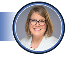
Kristy Crow, BS, RDCS, RDMS, RVT
Senior Sonographer / Radiation Sciences Educator
University of Iowa
Kristy Crow is a seasoned ultrasound professional with nearly 15 years of diverse experience. A proud alumna of the University of Iowa's Diagnostic Medical Sonography program, she has honed her skills across various echocardiography specialties, including adult congenital, pediatric, and fetal realms. Currently serving as a senior sonographer in pediatric cardiology, Kristy plays a pivotal role in diagnosing complex cardiac conditions in young patients and providing education and training to cardiology fellows. Beyond her clinical duties, she shares her wealth of knowledge as a Radiation Sciences instructor, nurturing aspiring sonographers. Kristy's dedication extends to volunteer work with prominent organizations like SDMS and ARDMS, underscoring her commitment to advancing the profession. Additionally, she lends her expertise as a consultant for Lantheus Imaging (Definity), ensuring optimal diagnostic accuracy through education and image optimization services. Kristy's multifaceted contributions epitomize her passion for excellence and innovation in medical imaging.
OB/GYN Track | 1.0 SDMS CME Credit | Category: OB | Content Level: Advanced
Gain an understanding of deep pelvic endometriosis (DPE) with a panel discussion. Presenters will discuss their experiences developing dedicated sonographic imaging for DPE for a large academic institution. A quick review of normal pelvic anatomy as well as interesting DPE cases involving bowel invasion and ligaments will be included.
Objectives:
- Review normal pelvic and bowel sonographic anatomy.
- Recognize common endometriosis/endometrioma sonographic findings and identify complex deep pelvis endometriosis invading the rectum/bowel and ligaments.
- Learn how to create protocols for your institution and identify available treatment options and surgical techniques.

Nadia Chupka, BS, RDMS, RVT
Sonographer
Mayo Clinic
Nadia M. Chupka has worked as a staff sonographer at Mayo Clinic in Rochester, MN for six years. She earned her Bachelor of Science degree in neuroscience from Michigan State University. She then went on to pursue sonography at the Mayo Clinic School of Health Sciences. She is registered through the American Registry for Diagnostic Medical Sonography holding credentials in Vascular, OB/Gyn, Abdominal, and Pediatric sonography. Nadia was the 2021 Society of Diagnostic Medical Sonography (SDMS) Emerging Leaders Grant recipient and currently enjoys serving on the SDMS Event Management Committee (EMC). She has presented at an SDMS Virtual Seminar, SDMS Knowledge Series event, and at the 2023 SDMS Annual Conference. Nadia has been recognized at work as the Clinical Instructor of the Year and a Silver Quality Fellow. She enjoys her role as the Wellness Champion of her department and is an advocate for enhancing the patient experience. Nadia has achieved the academic rank of Instructor and is a published author in the Journal of Diagnostic Medical Sonography (JDMS).

Talisha Hunt, BSRT, RDMS, RVT, RDCS, FSDMS
Lead Sonographer
Mayo Clinic
Talisha Hunt is a lead sonographer and Assistant Professor at Mayo Clinic in Rochester, MN and has been in the clinical practice for 19 years. She graduated from The University of Oklahoma Health Sciences Center and holds a Bachelor of Science Degree of Radiologic Technology in Sonography. Talisha is registered through ARDMS in the specialties of Abdomen, OB/GYN, Vascular, Adult Echocardiography and Pediatric Sonography. She considers volunteering as a way to give back to the profession. In addition to her service on the SDMS Board of Directors for seven years, Talisha is active within other SDMS committees and task forces, as well as a manuscript reviewer for the Journal of Diagnostic Medical Sonography and a member of the Editorial Board. Talisha has been an invited lecturer for several SDMS annual conferences and has taken home first and second place awards for the sonographer poster competition in previous years. She also has been an item writer for the ARDMS Pediatric Sonography examination and is a published author for multiple medical journal articles and two book chapters on liver, renal and pancreas transplantation.

Wendaline VanBuren, MD
Radiologist, Associate Professor of Radiology
Mayo Clinic
Dr. VanBuren is a board-certified radiologist, subspecialized in ultrasound, abdominopelvic and breast imaging. Dr. VanBuren is the chair of the Gynecological imaging section, with a dedicated interest in Endometriosis. Dr. VanBuren is one of the founding Directors for the Mayo Clinic Gynecological and Breast Imaging course. On a national level, she has co-founded and chaired the Disease Focused Panel on Endometriosis for the Society of Abdominal Radiology and was the invited guest editor for the journal Abdominal Radiology Special Edition on Endometriosis. Her ongoing work is frequently highlighted at a variety of national and international radiology and gynecology conferences. Dr. VanBuren takes pride in advocacy and propagating a passion for medical imaging, education, clinical and scientific collaboration.
Vascular Track | 1.0 SDMS CME Credit | Category: VT | Content Level: Beginner
This course will review anatomy and hemodynamics of the vertebral arteries. Waveform changes will also be discussed. Although the flow may be antegrade, is it normal? What is the waveform really trying to tell you?
Objectives:
- Review posterior cerebral anatomy and circulation.
- Discuss hemodynamic changes that can affect vertebral waveforms.
- Recognize abnormal vertebral waveforms and what they suggest.

Cathie Scholl, DHSc, RDMS, RVT, FSDMS
Associate Professor
Nova Southeastern University – Tampa Bay Regional Campus
Cathie Scholl is an associate professor in the Cardiovascular Sonography program at Nova Southeastern University-Tampa Bay regional campus. She earned a Doctor of Health Science degree in 2017 and was recognized as an SDMS Fellow in 2023. Cathie is a long-time volunteer for both the Society of Diagnostic Medical Sonography (SDMS) and the American Registry for Diagnostic Medical Sonography (ARDMS) and has served on many committees for both organizations. Currently, she is the chair of the ARDMS’ Sonographic Principles and Instrumentation exam. She is an associate editor for the Journal of Diagnostic Medical Sonography (JDMS) and serves on the editorial board. Cathie has authored original research and a case study, both published in the JDMS. In addition, she has contributed to two textbooks. Cathie teaches full-time and scans part-time.
General Session | 1.0 SDMS CME Credit | Category: OT | Content Level: Beginner
Stephen McLaughlin Memorial Lecture
In this presentation, I'll share my journey to securing an official Advanced Practice Sonographer position and offer tips for those aspiring to do the same. Additionally, I'll discuss the importance of formalizing Advanced Practice as a career progression option.
Objectives:
- Review the history of Advanced Practice and the important role that the SDMS played in its development.
- Share my personal journey towards becoming an Advanced Ultrasound Practitioner and provide characteristics that I believe are necessary for those that would like to pursue it.
- Summarize why I firmly believe that creating an official new Advanced Practice rung on the career ladder for the sonographer is critically important.

Ted Whitten, BA, RDMS, RVT, FSDMS
Advanced Ultrasound Practitioner
Elliot Hospital
With 30+ years in sonography,
Ted Whitten, BA, RDMS, RVT, FSDMS, past president of the Society of Diagnostic Medical Sonography (SDMS) and SDMS Foundation (2019-2021), continually fosters a dedication to helping others and to learning, which has helped him work in an advanced capacity at Elliot Hospital in Manchester, New Hampshire since 2007. Ted has elevated and standardized the quality of sonographic procedures for the Elliot Health system, served as a vital conduit between sonographers and physicians, travelled the U.S. on behalf of Elliot Hospital to teach sonography departments, and initiated a musculoskeletal sonography program at Elliot Hospital. At a prior employer, he established a non-invasive vascular lab that won accreditation by the Intersocietal Commission on Accreditation of Vascular Laboratories (ICAVL). Ted keeps his passion high through clinical connections with sonography students.
Morning Yoga | 0.75 SDMS CME Credit | Category: OT | Content Level: Beginner
Start your day with a 45-minute class that combines didactic information and movement. You'll learn stretching techniques to implement throughout your busy workday, helping to reduce stress and prevent musculoskeletal (MSK) injuries. This class is suitable for all levels, and mats will be provided. Comfortable clothing is recommended.
Objectives:
- Discuss the muscles of the neck and back including common areas of stress in the Sonographer.
- Learn effective tools to form habits throughout your day for overall wellness.
- Understand safe and effective stretching techniques to reduce tension throughout the body including the neck, back and shoulders.
Space is limited. Spots are available on a first come, first served basis.

Brandy Sundberg, MHPTT, RT (R), RDMS
Assistant Professor and Women’s Health Education Leader
University of Nebraska Medical Center and GE HealthCare
Brandy Sundberg is an Assistant Professor for the University of Nebraska Medical Center and Women's Health Education Leader for GE Healthcare. She has 20 years’ experience in Maternal Fetal Medicine where she was the Lead Sonographer, Sonographer Educator and Ultrasound Practitioner. Brandy graduated from the Diagnostic Medical Sonography program at the University of Nebraska Medical Center in 2002 with a Bachelor of Science in Radiation Science. She holds a Master’s Degree in Health Professions Teaching and Technology from the University of Nebraska Medical Center. Brandy is a Yoga Medicine trained yoga teacher and mindfulness meditation teacher. Brandy works with elite military professionals, healthcare professionals and individuals in her community to navigate stress and reduce burnout. Brandy has contributed to The Essential Guide to Yoga, focusing on the benefits of a restorative yoga practice. She is also an invited lecturer, discussing the benefits of mindfulness and stress reduction techniques for healthcare professionals, with a specific focus on alleviating musculoskeletal (MSK) injuries and burnout for sonographers. Brandy is co-founder of calmstrong.org, a website that provides functional movement and stress relief- created for healthcare professionals to integrate wellness into even the busiest of days.
Abdominal + Track | 1.0 SDMS CME Credit | Category: PS | Content Level: Intermediate
Join us for an insightful presentation delving into the realm of rare and unusual pathology observed in pediatric ultrasound cases; we will explore a diverse spectrum of pathology that can manifest in pediatric patients.
Objectives:
- Interactive discussion regarding not so common pediatric pathologies.
- Interesting cases to include a. Head/neck b. Body c. Extremities
- Illustrate the sonographic findings and their correlation with other modalities, as appropriate.

Tara Cielma, BS, RT(S), RDMS, RDCS, RVT, FSDMS
Sonography Research Specialist
Children's National Hospital
With over 16 years of experience, Tara is a well-established leader in the sonography field. Her credentials include abdomen, OB/GYN, neurosonography, vascular technology, fetal echo, and a credential from ARRT in Sonography. She is also designated as an SDMS Fellow.
Tara's dedication to advancing the field of sonography extends beyond her professional roles; she is deeply committed to giving back to the sonography community. Tara actively volunteers with SDMS, ARDMS/Inteleos, and SPR (just to name a few). She also reviews manuscripts for the Journal of Diagnostic Medical Sonography (JDMS), contributes to writing registry items and preparation material, and provides hands-on workshops for facilities seeking to develop or enhance their pediatric and fetal imaging skillset.
Tara has numerous chapters, journal articles, and a co-authored pediatric registry review book to her name. Tara's extensive experience, unwavering dedication, and passion for volunteerism ensure she will be making waves in the field for years to come.

Monique Riemann, RDMS, RVT
Research Sonographer
Phoenix Children’s Hospital
Monique Riemann is an ultrasound technologist with 37 years of experience in the field. Her registries include Abdomen, OB/GYN, Vascular, (Neuro), and Pediatric. She began her career at Jackson Memorial Medical Center in Miami, Florida, before relocating to Arizona. After many years in a lead position at Phoenix Children’s Hospital, she transitioned to a research position where she has played a significant role in creating ultrasound exams that were once radiation-based, utilizing the motto "Think Ultrasound First." Monique has received awards for scientific presentations and poster competitions at the Society of Diagnostic Medical Sonography (SDMS). Over the last 7 years, she has had the pleasure of speaking at several conferences. To date, she has over 20 publications on various topics related to ultrasound. Monique's passion for ultrasound extends beyond her research role; she is also employed part-time as an educator and lab instructor at West Coast Ultrasound Institute, where she contributes to the development of future sonographers.
Cardiac Track | 1.0 SDMS CME Credit | Category: AE | Content Level: Intermediate
This presentation will include friendly competition, while reviewing fun and interesting echocardiograms.
Objectives:
- Have Fun.
- Engage in Friendly competition.
- Win and go home with goodies.

Neha Soni-Patel, MEd, BSME, RDCS (AE/PE), RCCS, FASE
Education Work Leader
Cleveland Clinic Children’s
Neha is a senior sonographer and Education Work Leader at Cleveland Clinic Children's. She holds a Master of Education for Health Professionals from Cleveland State University and a bachelor’s in mechanical engineering from Purdue University. Neha is an instructor of Pediatric Echo at Cuyahoga Community College. A long-time SDMS member, she educates new sonographers on Congenital Heart Defects (CHD) and their transition into adulthood as well as how to approach undiagnosed congenital heart defects. Neha recently contributed to new Pediatric Echo Guidelines published in the Journal of the American Society of Echocardiography and serves on its board of directors. She also serves as an SDMS CME reviewer and on the editorial board of the JDMS. Passionate about education, Neha believes in the power of lifelong learning.
OB/GYN Track | 1.0 SDMS CME Credit | Category: FE | Content Level: Advanced
Discover essential techniques for fetal cardiac screening, recognize common congenital heart defects, and identify fetal arrhythmias in this comprehensive lecture. Gain proficiency in acquiring fetal views, understanding common congenital heart defects like Tetralogy of Fallot, and recognizing arrhythmias such as supraventricular tachycardia. This lecture is perfect for healthcare professionals seeking to enhance prenatal cardiac evaluation skills.
Objectives:
- Identify basic fetal echo views for screening congenital heart defects.
- Differentiate common congenital heart defects and fetal echo views for diagnosis.
- Evaluate and identify fetal arrhythmias using echocardiography.

Kristy Crow, BS, RDCS, RDMS, RVT
Senior Sonographer / Radiation Sciences Educator
University of Iowa
Kristy Crow is a seasoned ultrasound professional with nearly 15 years of diverse experience. A proud alumna of the University of Iowa's Diagnostic Medical Sonography program, she has honed her skills across various echocardiography specialties, including adult congenital, pediatric, and fetal realms. Currently serving as a senior sonographer in pediatric cardiology, Kristy plays a pivotal role in diagnosing complex cardiac conditions in young patients and providing education and training to cardiology fellows. Beyond her clinical duties, she shares her wealth of knowledge as a Radiation Sciences instructor, nurturing aspiring sonographers. Kristy's dedication extends to volunteer work with prominent organizations like SDMS and ARDMS, underscoring her commitment to advancing the profession. Additionally, she lends her expertise as a consultant for Lantheus Imaging (Definity), ensuring optimal diagnostic accuracy through education and image optimization services. Kristy's multifaceted contributions epitomize her passion for excellence and innovation in medical imaging.
Vascular Track | 1.0 SDMS CME Credit | Category: VT | Content Level: Intermediate
This presentation will discuss the anatomy, physiology, and pathophysiology associated with the unique vascular system that serves the liver.
Objectives:
- Discuss vascular anatomy and hemodynamics related to hepatoportal blood flow.
- Discuss the physiology and pathophysiology related to hepatoportal blood flow.
- Identify imaging protocols to properly assess hepatoportal blood flow.
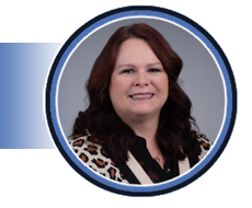
Katherine Peak, EdD, RT(R), RDMS, RVT
Clinical Assistant Professor
University of Southern Indiana
Katherine Peak has been a sonographer for the past 27 years. Her career has included working in a busy sonography department in an acute care hospital and as the technical director of a vascular lab for a large group of vascular surgeons. For the past 12 years, Katherine has had the privilege of teaching general and vascular sonography students at the University of Southern Indiana and serving as their clinical coordinator. She is very involved with her professional societies, where she currently serves the SDMS on the event management committee and the nominating committee. Katherine is also the sonography chapter delegate to the ASRT House of Delegates and is the sonography representative on the ASRT Practice Standards council.
Educator Track | 1.0 SDMS CME Credit | Category: OT | Content Level: Beginner
Accreditation requires facilities to meet minimum standards for quality patient care. Compliance with these standards reduces risk management issues through care standardization and mandatory self-assessment for continuous improvement.
Objectives:
- Introduce and explain the value of complying with minimum testing Standards to improve and maintain quality patient care.
- Compliance with the Standards by all staff will ensure a Standard of care that may help to mitigate risk management issues.
- Accreditation requires continuous self-improvement reviews to ensure all staff are maintaining compliance with the Standards.
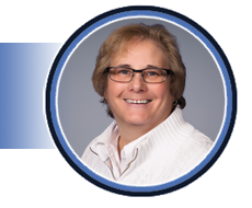
Marge Hutchisson, BS, RVT, RDCS, RPhS
Director
IAC
Marge Hutchisson, BS, RVT, RDCS, RPhS joined the IAC in April 2008 and serves as the Director of Accreditation for IAC Vascular Testing. Her experience includes more than 30 years in the field of peripheral vascular ultrasound and 13 years in transthoracic echocardiography. Throughout her career, she has served in a managerial and technical director capacity in the establishment and management of cardiovascular laboratories. Ms. Hutchisson earned her nursing diploma at Lakewood School of Practical Nursing in Cleveland, Ohio, and her Bachelor of Science degree from Colorado State University in Fort Collins, CO. She is board-certified by the American Registry of Diagnostic Medical Sonography (ARDMS) in both Vascular Technology and Adult Echocardiography. She is credentialed by Cardiovascular Credentialing International (CCI) as a Registered Phlebology Sonographer. She is an active member of the ARDMS, Society of Vascular Ultrasound (SVU) and CCI.
Abdominal + Track | 1.0 SDMS CME Credit | Category: AB | Content Level: Inermediate
Appendicitis is an acute abdominal emergency that can occur in patients of all ages. Ultrasound is a reliable tool in the diagnosis of appendicitis and differential potentials. Due to its lack of radiation, ultrasound should be considered as a first line of evaluation in the detection of acute appendicitis. This presentation will focus on anatomy of the appendix, normal versus abnormal sonographic features of the appendix, as well as primary and differential diagnosis potentials.
Objectives:
- Understand the anatomy and physiology of the appendix.
- Discuss scan technique in bowel imaging.
- Distinguish normal from abnormal findings encountered during appendix evaluation.

Patricia Lacy Gandor, MS, RDMS, RVT, RMSKS
GE HealthCare
Clinical Applications Specialist, Point of Care Ultrasound
Patricia Lacy Gandor is an assistant professor and clinical coordinator of sonography programs at Harper College. She graduated from Southern Illinois University with a bachelor’s degree in allied health; radiography and sonography, earned a master’s degree in health profession education from Rutgers University, and is now pursuing doctoral studies in curriculum and instruction at Northern Illinois University. Lacy is credentialed through the American Registry for Diagnostic Medical Sonography (ARDMS) in abdomen, OB/GYN, breast, pediatric, vascular, and musculoskeletal imaging. With over 20 years of clinical and educational experience, she is passionate about sonography and collaborating with others on global health, research, and education. In her spare time, Lacy enjoys traveling, boating, hiking, and golf.
Cardiac Track | 1.0 SDMS CME Credit | Category: AE | Content Level: Beginner
Echocardiography is one of the most technically dependent non-invasive imaging modalities. Performing an echocardiogram requires the sonographer to be passionate about their role in image acquisition and analysis. Proper image acquisition and accurate, reproducible measurements are necessary to make the correct diagnosis and determine treatment. A small variation in a single measurement can impact calculations of valve areas, regurgitation grades, etc. An improper angel can miss an abnormal flow. Failure to be thorough can miss a thrombus or wall motion abnormality. Not attempting to adequately answer the clinical question impacts the quality of care and can lead to further, unnecessary medical testing. Utilizing cases studies, this presentation will demonstrate the importance of precision and the role that passion plays in performing quality echocardiograms.
Objectives:
- Explain how an improper measurement impacts the calculation of valve areas and regurgitation gradients.
- Describe the role of proper image acquisition in the assessment of cardiac size and function.
- Discuss how the passion, persistence, and intentionality of the sonographer impact the quality of the information provided by the echocardiogram.
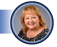
Elizabeth McIlwain, MHS, ACS, RCS, FASE
Clinical Director Procedural/Outpatient Services
West Jefferson Medical Center / LCMC Health
Elizabeth is a graduate of Louisiana State University Health Sciences Center, holding a Bachelor of Science degree in Cardiopulmonary Science and a Master of Health Science degree in Health Care Administration. She is a registered cardiac sonographer (RCS) and an advanced cardiac sonographer (ACS), with a career spanning since 1985 in the field of cardiovascular ultrasound. Actively involved in the profession at both local and national levels, Elizabeth promotes cardiovascular sonography locally through participation in educational events and as a resource to sonographers in the New Orleans area. At the national level, she volunteers for organizations such as the American Society of Echocardiography, Cardiovascular Credentialing International, and the Society of Diagnostic Medical Sonography, serving in various leadership capacities. Elizabeth has contributed significantly to research, with multiple publications to her credit and co-authorship of ‘The Handbook of Echo Doppler Interpretation.’ Her work experience is diverse, having served in roles such as staff sonographer, lead sonographer, researcher, educator, industry consultant, and department director. Currently, Elizabeth holds the position of Clinical Director of Procedural/Outpatient Services at West Jefferson Medical Center in Marrero, LA, providing leadership in Cardiology, Special Procedures, Cardiac & Pulmonary Rehab, Endoscopy, Hyperbarics/Wound Care, and Dialysis. Additionally, she serves as clinical faculty in the cardiovascular ultrasound program at LSUHSC in New Orleans. Passionate about "all things echo," Elizabeth frequently describes herself as "just an echo nerd."
OB/GYN Track | 1.0 SDMS CME Credit | Category: OB | Content Level: Intermediate
This presentation delves into the fascinating world of obstetric imaging, showcasing compelling case studies that demonstrate the efficacy of employing multiple modalities in prenatal care. We will explore how combining ultrasound, MRI, and other imaging techniques can enhance our understanding of fetal development, diagnose complex conditions, and guide clinical decision-making. Through detailed case presentations, we will highlight the strengths and limitations of each modality, discuss best practices for integration, and showcase the benefits of a multimodal approach in obstetric imaging.
Objectives:
- Briefly review imaging modalities utilized in obstetric cases and their limitations concerning fetal imaging.
- Review obstetric case studies that utilize multiple imaging modalities to aid in the diagnosis and management of fetal anomalies.
- Discuss collaborative approaches to managing these cases prenatally and postnatally.

Kelsi McConnell, MHS, RDMS, RVT
Practice Manager
Regional One Health
Kelsi McConnell is an accomplished Practice Manager specializing in Maternal Fetal Medicine (MFM), with a wealth of experience in ultrasound training and education. She has managed clinical practices and worked as an MFM sonographer for the last 9 years. Kelsi is an adjunct professor at Baptist University of Health Sciences and serves on the advisory board of the Diagnostic Medical Sonography (DMS) program. She earned her Bachelor’s Degree in Diagnostic Medical Sonography from Baptist University of Health Sciences and went on to obtain her Master’s Degree in Leadership and Policy Studies from the University of Memphis. Additionally, she is a contributing author of two chapters in the Textbook of Diagnostic Sonography (9th ed.) titled “Prenatal Diagnosis of Congenital Anomalies” and “Fetal Skeleton”. Kelsi serves as a JRC-DMS Site Visitor and has been heavily involved in the SDMS, having served on several committees for the organization over the past 10+ years.
Vascular Track | 1.0 SDMS CME Credit | Category: VT | Content Level: Beginner
This presentation will feature case studies covering peripheral vascular, abdominal, obstetric, and gynecologic sonography. It will discuss patient presentation, disease etiology, the role of sonography in diagnosis, and treatment outcomes.
Objectives:
- Identify vascular pathologies based on patient presentation and risk factors.
- Summarize sonographic findings associated with vascular pathologies.
- Discuss potential patient outcomes of vascular pathologies.

Jennifer Bagley, MPH, RDMS, RVT, FAIUM, FSDMS
Professor, Program Director
The University of Oklahoma Health Sciences Center
Jennifer Bagley, MPH, RDMS, RVT is a Professor and Sonography Program Director at The University of Oklahoma Health Sciences Center. Jennifer has maintained SDMS membership for nearly 30 years and served on the Board of Directors for 4 years. She has participated in various SDMS committees including the Continuing Education Committee, Nominating Committee, Conference Management Committee, and the Research Institute Task Force. She has served as the SDMS Liaison to the AIUM Bioeffects and Safety Committee since 2009. Jennifer is a past winner of the SDMS Distinguished Educator Award as well as a past first and second place winner of the Journal of Diagnostic Medical Sonography’s Kenneth R. Gottesfeld Award.
Educator Track | 1.0 SDMS CME Credit | Category: OT | Content Level: Intermediate
Most health professions programs, including sonography, have a clinical education component. It is in this learning environment that students move theory to practice and learn how to become a professional in their field of study. Belongingness, the feeling of being accepted or connected to others in the clinical environment, improves student motivation to learn, self-confidence, and skills competence. In contrast, the feeling of isolation in this learning environment has been associated with increased student stress and anxiety and reduced motivation to learn. This presentation will cover novel information gathered from a quantitative study that explored diagnostic medical sonography (DMS) students’ feeling of belongingness in the clinical learning environment. Study findings may help educational programs address belongingness with their clinical sites and improve the DMS student learning experience.
Objectives:
- Define belongingness as a universal human need.
- Describe how belongingness or the lack of belongingness in the clinical setting affects health professions students’ learning.
- Translate how the findings from this study can be used to improve the clinical learning environment for sonography students.
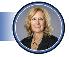
Kimberly Michael, EdD, MA, RT(R), RDMS, RVT, FSDMS
Director, Diagnostic Medical Sonography Education
University of Nebraska Medical Center
Kim Michael, EdD, RDMS, RVT, FSDMS, is a Professor in the College of Allied Health Professions at the University of Nebraska Medical Center (UNMC) where she also holds the Anderson Distinguished Professorship. She has been involved in sonography education at UNMC since 1991 and has served as the Director of the Diagnostic Medical Sonography program since 1998. She received her Bachelor of Science Degree from UNMC, a Master of Arts degree from the University of Nebraska at Omaha and a Doctor of Education from Clarkson College. Kim has authored or co-authored over 50 publications related to sonography and health professions education. Her current research interests include interprofessional education and belongingness in the clinical learning environment.
Abdominal + Track | 1.0 SDMS CME Credit | Category: AB | Content Level: Intermediate
In this presentation, we'll explore uncommon pediatric pathologies that often leave us wondering 'What is that?!'. We'll share tips to aid in identifying these conditions and review essential information for rendering a definitive diagnosis.
Objectives:
- Explore uncommon pathologies that can be encountered in the pediatric patient.
- Learn some helpful tips that will help us answer the question, 'What is that?!'.
- Summarize the important information needed to help render a definitive diagnosis.

Ted Whitten, BA, RDMS, RVT, FSDMS
Advanced Ultrasound Practitioner
Elliot Hospital
With 30+ years in sonography,
Ted Whitten, BA, RDMS, RVT, FSDMS, past president of the Society of Diagnostic Medical Sonography (SDMS) and SDMS Foundation (2019-2021), continually fosters a dedication to helping others and to learning, which has helped him work in an advanced capacity at Elliot Hospital in Manchester, New Hampshire since 2007. Ted has elevated and standardized the quality of sonographic procedures for the Elliot Health system, served as a vital conduit between sonographers and physicians, travelled the U.S. on behalf of Elliot Hospital to teach sonography departments, and initiated a musculoskeletal sonography program at Elliot Hospital. At a prior employer, he established a non-invasive vascular lab that won accreditation by the Intersocietal Commission on Accreditation of Vascular Laboratories (ICAVL). Ted keeps his passion high through clinical connections with sonography students.
Cardiac Track | 1.0 SDMS CME Credit | Category: AE | Content Level: Intermediate
This presentation will cover adding different views to the adult echo protocol to help identify congenital heart defects. The presenter will discuss common pitfalls when encountering a Congenital Heart Defect (CHD.)
Objectives:
- Review the adult protocol with addition of pediatric views.
- Understand simple defects that can be seen in a routine adult echo.
- Review undiagnosed or unknown CHDs.

Neha Soni-Patel, Med, BSME, RDCS (AE/PE), RCCS, FASE
Education Work Leader
Cleveland Clinic Children’s
Neha is a senior sonographer and Education Work Leader at Cleveland Clinic Children's. She holds a Master of Education for Health Professionals from Cleveland State University and a bachelor’s in mechanical engineering from Purdue University. Neha is an instructor of Pediatric Echo at Cuyahoga Community College. A long-time SDMS member, she educates new sonographers on Congenital Heart Defects (CHD) and their transition into adulthood as well as how to approach undiagnosed congenital heart defects. Neha recently contributed to new Pediatric Echo Guidelines published in the Journal of the American Society of Echocardiography and serves on its board of directors. She also serves as an SDMS CME reviewer and on the editorial board of the JDMS. Passionate about education, Neha believes in the power of lifelong learning.
OB/GYN Track | 1.0 SDMS CME Credit | Category: OB | Content Level: Intermediate
This lecture will provide a comprehensive overview of both benign and malignant ovarian pathology, including discussions on physiologic processes, ovarian torsion, and polycystic ovarian syndrome. Additionally, we'll explore sonographic techniques and strategies to enhance scanning for accurate diagnosis. We'll also address when intervention is warranted and when it may not be required.
Objectives:
- Recognize benign vs malignant sonographic characteristics of ovarian masses.
- Identify scanning techniques to elicit more sonographic information to aid in diagnosis.
- Understand when intervention may be necessary.

Nadia Chupka, BS, RDMS, RVT
Sonographer
Mayo Clinic
Nadia M. Chupka has worked as a staff sonographer at Mayo Clinic in Rochester, MN for six years. She earned her Bachelor of Science degree in neuroscience from Michigan State University. She then went on to pursue sonography at the Mayo Clinic School of Health Sciences. She is registered through the American Registry for Diagnostic Medical Sonography holding credentials in Vascular, OB/Gyn, Abdominal, and Pediatric sonography. Nadia was the 2021 Society of Diagnostic Medical Sonography (SDMS) Emerging Leaders Grant recipient and currently enjoys serving on the SDMS Event Management Committee (EMC). She has presented at an SDMS Virtual Seminar, SDMS Knowledge Series event, and at the 2023 SDMS Annual Conference. Nadia has been recognized at work as the Clinical Instructor of the Year and a Silver Quality Fellow. She enjoys her role as the Wellness Champion of her department and is an advocate for enhancing the patient experience. Nadia has achieved the academic rank of Instructor and is a published author in the Journal of Diagnostic Medical Sonography (JDMS).
Vascular Track | 1.0 SDMS CME Credit | Category: VT | Content Level: Intermediate
Internal carotid artery stenosis interpretation has become a significant public health issue as many facilities use different, non-validated criteria. This practice has left the patient with an uncertain, possibly inaccurate percent stenosis.
Objectives:
- Review the need for standardization across all facilities to use the same diagnostic criteria for internal carotid artery stenosis.
- Explain the need to adopt standardized, validated criteria to ensure accurate interpretation of internal carotid artery stenosis.
- Continuous quality improvement activities will ensure that all staff are adhering to the facility interpretation criteria.

Marge Hutchisson, BS, RVT, RDCS, RPhS
Director
IAC
Marge Hutchisson, BS, RVT, RDCS, RPhS joined the IAC in April 2008 and serves as the Director of Accreditation for IAC Vascular Testing. Her experience includes more than 30 years in the field of peripheral vascular ultrasound and 13 years in transthoracic echocardiography. Throughout her career, she has served in a managerial and technical director capacity in the establishment and management of cardiovascular laboratories. Ms. Hutchisson earned her nursing diploma at Lakewood School of Practical Nursing in Cleveland, Ohio, and her Bachelor of Science degree from Colorado State University in Fort Collins, CO. She is board-certified by the American Registry of Diagnostic Medical Sonography (ARDMS) in both Vascular Technology and Adult Echocardiography. She is credentialed by Cardiovascular Credentialing International (CCI) as a Registered Phlebology Sonographer. She is an active member of the ARDMS, Society of Vascular Ultrasound (SVU) and CCI.
Educator Track | 1.0 SDMS CME Credit | Category: OT | Content Level: Beginner
This presentation will explore how ultrasound educators can begin conducting and sharing research. It will cover topics like selecting a research topic and navigating the institutional review board process. Additionally, it will provide insights on disseminating research and scholarship.
Objectives:
- Explain the importance of research for ultrasound educators.
- Illustrate research projects that are feasible for ultrasound educators.
- Identify the steps the ultrasound educator needs to take a project from an idea to disseminated scholarship.

Jennifer Bagley, MPH, RDMS, RVT, FAIUM, FSDMS
Professor, Program Director
The University of Oklahoma Health Sciences Center
Jennifer Bagley, MPH, RDMS, RVT is a Professor and Sonography Program Director at The University of Oklahoma Health Sciences Center. Jennifer has maintained SDMS membership for nearly 30 years and served on the Board of Directors for 4 years. She has participated in various SDMS committees including the Continuing Education Committee, Nominating Committee, Conference Management Committee, and the Research Institute Task Force. She has served as the SDMS Liaison to the AIUM Bioeffects and Safety Committee since 2009. Jennifer is a past winner of the SDMS Distinguished Educator Award as well as a past first and second place winner of the Journal of Diagnostic Medical Sonography’s Kenneth R. Gottesfeld Award.
Abdominal + Track | 1.0 SDMS CME Credit | Category: AB | Content Level: Beginner
Submerge yourself into basic and interesting emergency department sonographic cases. This session will keep you engaged and test your knowledge. While some cases may be a review, others require critical thinking. Confirm what you already know and learn a few things along the journey.
Objectives:
- Identify normal sonographic anatomy and recognize and identify common and uncommon sonographic pathology within the emergent setting.
- Identify appropriate imaging and/or correlative examinations to help aid in accurate diagnosis.
- Identify available treatment options.

Talisha Hunt, BSRT, RDMS, RVT, RDCS, FSDMS
Lead Sonographer
Mayo Clinic
Talisha Hunt is a lead sonographer and Assistant Professor at Mayo Clinic in Rochester, MN and has been in the clinical practice for 19 years. She graduated from The University of Oklahoma Health Sciences Center and holds a Bachelor of Science Degree of Radiologic Technology in Sonography. Talisha is registered through ARDMS in the specialties of Abdomen, OB/GYN, Vascular, Adult Echocardiography and Pediatric Sonography. She considers volunteering as a way to give back to the profession. In addition to her service on the SDMS Board of Directors for seven years, Talisha is active within other SDMS committees and task forces, as well as a manuscript reviewer for the Journal of Diagnostic Medical Sonography and a member of the Editorial Board. Talisha has been an invited lecturer for several SDMS annual conferences and has taken home first and second place awards for the sonographer poster competition in previous years. She also has been an item writer for the ARDMS Pediatric Sonography examination and is a published author for multiple medical journal articles and two book chapters on liver, renal and pancreas transplantation.
Cardiac Track | 1.0 SDMS CME Credit | Category: AE | Content Level: Intermediate
Aortic stenosis is a common pathology encountered by echocardiographers. Its various causes and appearances make it challenging to diagnose accurately. This presentation will address scanning factors, patient demographics, and barriers associated with this condition.
Objectives:
- Understand the basics of working up aortic stenosis from a transthoracic echo standpoint.
- Learn the different causes and appearances of aortic stenosis.
- Feel confident that they understand the hemodynamics involved in this complex pathology.
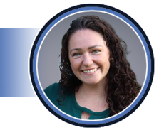
Jaclyn Meadow, RCS
Senior Ultrasound Clinical Account Specialist
Biosense Webster
Jaclyn Meadow, Senior Ultrasound Clinical Account Specialist at Biosense Webster, specializes in guiding physicians through intracardiac echocardiography in electrophysiology and structural heart interventions. Her journey started with formal training at the Sanford Brown Institute in Tampa, Florida, and flourished during her tenure at Orlando Regional Medical Center. With a focus on structural heart cases and advanced pathology, Jaclyn mastered cutting-edge 3D imaging and quantification techniques over her 12-year career. Now at Biosense Webster, she shares her expertise to help healthcare professionals navigate intracardiac imaging complexities, contributing to the continuous advancement of echocardiography.
OB/GYN Track | 1.0 SDMS CME Credit | Category: FE | Content Level: Advanced
Congenital heart disease is present in 60-80% of patients with DiGeorge Syndrome and the most frequently seen cardiac malformations are Conotruncal defects. This presentation will discuss and review imaging goals and tips for congenital heart disease associated with DiGeorge Syndrome.
Objectives:
- Review DiGeorge Syndrome.
- Discuss lesion specific scanning goals.
- Review fetal echo images for conotruncal defects.
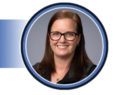
Dawn Park, BS, RDCS, RCCS, FASE
Pediatric Cardiac Sonographer Educator
Nationwide Children’s Hospital
Dawn Park launched her career as a cardiac sonographer in Michigan in 2000, following her graduation from the College of West Virginia. While initially focused on scanning adult patients, she seized the opportunity to learn pediatric echo, discovering her passion for congenital heart disease. In 2009, she transitioned to Children’s Hospital Colorado as a pediatric and fetal cardiac sonographer and education coordinator. After 13 years in Colorado, she traded mountains for beaches, enjoying what she calls 'a very long vacation' in Florida. Recently, Dawn joined Nationwide Children’s Hospital in Columbus, Ohio, as a pediatric cardiac sonographer educator. She also volunteers with ASE and the Society of Pediatric Echocardiography.
Vascular Track | 1.0 SDMS CME Credit | Category: VT | Content Level: Beginner
This presentation will discuss the proper scanning technique and different limitations sonographers may face when performing carotid duplex evaluations. Participants will learn to identify the common pitfalls when performing duplex and Doppler evaluations of carotid vessels. The session will provide practical tips and recommendations for best practices and best approaches when performing a carotid duplex examination.
Objectives:
- Describe patient limitations and proper scanning technique.
- Learn to identify the common pitfalls in carotid duplex evaluations.
- Discuss the importance of proper ultrasound instrumentation and anticipation of potential errors.

Patricia Vargas, DHSc, RVT
Professor & Program Director
Touro University
Patricia Vargas earned a Doctor of Health Science degree from A.T. Still University and a Master of Health Science from Nova Southeastern University. She serves as Professor and Program Director of the new DMS Program at Touro University, and she was as a Professor and Clinical Coordinator of the Medical Sonography Program at Nova Southeastern University prior to her appointment at Touro University. Dr. Vargas is a specialized vascular sonographer with over 18 years of experience in the field. Her dedication to education and her commitment to excellence are evident in her research interests, which include vascular sonography, diabetes, clinical education, sonography education, and interprofessional education. She is an active member of the Society of Diagnostic Medical Sonography (SDMS), the Society for Vascular Ultrasound (SVU), and the American Institute of Ultrasound in Medicine (AIUM). Her contributions as an invited speaker, subject matter expert, and peer reviewer reflect her passion for advancing the field of sonography.
Educators Track | 1.0 SDMS CME Credit | Category: OT | Content Level: Beginner
In this lively 50-minute talk, renowned clinical psychologist and author Dr. Monica Vermani shares her pragmatic and stigma-shattering approach to forging a deeper wellness by recognizing how symptoms manifest and play out, and understanding the causes and impacts of stress and burnout. She shares powerful and practical self-care tools to help manage life's stressors and an inspiring approach to imagining and creating the life you truly want.
Objectives:
- Attendees will learn Dr. Vermani’s patented ‘problems-on-the-table’ approach to identifying and addressing mental health issues, from mood disorders, traumas, substance abuse, and other problematic habits, behaviors, and choices that undermine and threaten our well-being, and creating a pathway to healing and growth.
- Audiences will gain a deep understanding of the complex and varied sources of their negative thoughts.
- Audiences will gain an in-depth awareness and understanding of how problems manifest and play out their lives.

Monica Vermani, C. Psych.
Clinical Psychologist, CEO and Founder
Dr. Vermani Balanced Wellbeing
Dr. Monica Vermani is a clinical psychologist specializing in treating trauma, stress, mood, and anxiety disorders, and a well-known public speaker, teacher, author, influencer, and mental health advocate. In over 25 years of clinical practice, Dr. Vermani has successfully treated thousands of patients through Supportive Psychotherapy, Cognitive Behavioral Therapy (CBT), Eye Movement Desensitization Reprocessing (EMDR), Breath~Body~Mind (BBM) practices, Mindfulness Meditation, Mindfulness-Based Stress Reduction (MBSR), and Mindfulness-Based Cognitive Therapy (MBCT). Dr. Vermani is a regular expert commentator/contributor to online and print media outlets, and TV, radio, and podcasts, including Forbes Magazine, CNN Healthline, Psychology Today, InStyle Magazine, Parade, HUFFPOST, Martha Stewart, Fast Company, Oprah Daily, and many others. Her latest book, A Deeper Wellness, conquering stress, mood anxiety, and traumas, is available in hard copy, eBook, and audiobook worldwide on Amazon.
Abdominal + Track | 1.0 SDMS CME Credit | Category: AB | Content Level: Intermediate
Ultrasound is a powerful imaging tool to evaluate the gallbladder and bile ducts. In this lecture, we will go over the details of the biliary tract and how it works, how we, as sonographers, can use anatomic, acoustic windows to better optimize images of this system, and we will go over some common and not-so common pathologies. We will also discuss how contrast enhanced ultrasound (CEUS) can be used to better confirm pathologies involving the biliary tract. We will end with a case review of the pathologies of the biliary tract to test our knowledge!
Objectives:
- Understand the normal anatomy and physiology of the biliary tract (gallbladder and bile ducts) and discuss the sonographic protocol and image optimization techniques to evaluate the biliary tract.
- Discuss the many pathologies that can encompass the biliary tract and the sonographic appearances of these pathologies.
- Understand how contrast enhanced ultrasound (CEUS) can be used to evaluate pathologies of the biliary tract.

Sara Baker, Med, RDMS, RVT, RMSKS, RT(R), FSDMS
Program Manager, US/NM/PET Accreditation
American College of Radiology
Sara has been a registered diagnostic medical sonographer and registered vascular technologist for over 20 years. Her sonography specialties include abdomen, vascular, obstetrics, gynecology, pediatric/neurosonography, and musculoskeletal. Sara is an active professional volunteer. She has been a site visitor for the JRC-DMS from 2012-2021, is an external reviewer, and is a virtual site visitor liaison. She also volunteers with the ARDMS and SDMS. She is the Chair of the Simulation Task force for Inteleos/ARDMS, was the Chair and Vice Chair for the Pediatric Specialty Exam Committee from 2019-2023, and she has written items for the ARDMS board exams in the areas of Pediatric, Abdomen, and Breast Sonography. In addition, she currently serves on the Board of Directors for the SDMS and the SDMS Foundation. She served in those roles for the SDMS and the SDMS Foundation from 2015-2017 and served on the Nominating Committee for the SDMS from 2017-2021. She has also given several webinars for the SDMS. Lastly, Sara has given lectures on a national level for the SDMS, on a local level for the South Central Wisconsin Ultrasound Society (SCWUS) and the Wisconsin Society of Radiologic Technologists (WSRT), and has submitted and collaborated with others in publishing research articles and textbook chapters.
Cardiac Track | 1.0 SDMS CME Credit | Category: AE | Content Level: Intermediate
Intracardiac Echocardiography (ICE) is now widely utilized in electrophysiology labs and is increasingly being adopted in catheterization (Cath) labs and structural heart procedures. As an echocardiographer, having a fundamental understanding of the various ICE catheters you may encounter, along with the images they produce, is essential. This presentation will cover the basic maneuvers of an ICE catheter, its movement within the heart, and the access points used by physicians during procedures.
Objectives:
- Understand the Fundamentals of ICE Catheters: Learn the types and imaging capabilities of ICE catheters used in labs.
- Master Basic Maneuvers and Navigation: Understand basic ICE catheter maneuvers for optimal imaging.
- Procedural Integration: Understand access points and how to integrate ICE imaging into cardiac procedures and their role as a sonographer.

Jaclyn Meadow, RCS
Senior Ultrasound Clinical Account Specialist
Biosense Webster
Jaclyn Meadow, Senior Ultrasound Clinical Account Specialist at Biosense Webster, specializes in guiding physicians through intracardiac echocardiography in electrophysiology and structural heart interventions. Her journey started with formal training at the Sanford Brown Institute in Tampa, Florida, and flourished during her tenure at Orlando Regional Medical Center. With a focus on structural heart cases and advanced pathology, Jaclyn mastered cutting-edge 3D imaging and quantification techniques over her 12-year career. Now at Biosense Webster, she shares her expertise to help healthcare professionals navigate intracardiac imaging complexities, contributing to the continuous advancement of echocardiography.
OB/GYN Track | 1.0 SDMS CME Credit | Category: OB | Content Level: Advanced
This presentation will review the types of diabetes seen during pregnancy, including Type 1, Type 2, and Gestational Diabetes. As sonographers, it is essential to understand and strive to correlate images with the clinical history of diabetes.
Objectives:
- Describe the congenital anomalies of the pregestational diabetic women that can be detected with an obstetrical ultrasound examination.
- Define the modified White classification used for pregnant diabetic patients.
- Explain the ultrasound measurement techniques which can be used to determine large for gestational age fetuses.
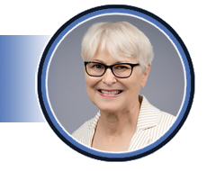
Pamela Foy, MS, RDMS, FSDMS, FAIUM
Imaging Training Program Manager
The Ohio State University
Pamela Foy currently serves as the Imaging Training Program Manager for the Department of Women’s Imaging and holds the position of Clinical Associate Professor in the Department of Obstetrics and Gynecology at The Ohio State University. With a distinguished career spanning 45 years, Pam is a fellow of both the Society of Diagnostic Medical Sonography (SDMS) and the American Institute of Ultrasound in Medicine (AIUM). Pam's educational background includes a certificate in diagnostic sonography from Thomas Jefferson University, a BS in Radiologic Technology from Widener University, and an MS from The Ohio State University. She is deeply passionate about sonography and has been actively involved in clinical sonography, education, and research throughout her career. In addition to her professional roles, Pam enjoys volunteering her time and expertise. She has served on various committees, including the SDMS Conference Management Committee and the Pediatric Sonography Examination for the American Registry for Diagnostic Medical Sonography (ARDMS). Pam was also a member of the board of governors for the AIUM. Pamela Foy has been sharing her knowledge and expertise in sonography since 1994 when she gave her first SDMS presentation. With her wealth of experience and dedication, Pam believes that sonography offers an excellent career path for those interested in the field.
Vascular Track | 1.0 SDMS CME Credit | Category: VT | Content Level: Beginner
This presentation delves into the invaluable role of ultrasound in diagnosing mesenteric artery disorders, including aneurysms, stenosis, and common compression syndromes. Starting with an overview of mesenteric anatomy and blood supply, it progresses to the principles of ultrasound imaging techniques tailored for detecting abnormalities in the mesenteric vasculature.
Objectives:
- Understand the anatomical landmarks and vascular supply of the mesenteric arteries.
- Differentiate between physiological and pathological mesenteric artery stenosis using ultrasound imaging criteria such as velocity measurements and spectral waveform analysis.
- Discuss the clinical significance of mesenteric artery pathologies diagnosed via ultrasound and their implications for patient management, including treatment options and potential complications.

Aubrey Rybyinski, BS, RDMS, RVT, FSDMS
Strategic Account Clinical and Operations Director
Navix Diagnostix
Aubrey Rybyinski, BS, RDMS, RVT, FSDMS, is the NAVIX Strategy Clinical and Operations Director based at Cooper University Department of Vascular Surgery, holding credentials from the American Registry for Diagnostic Medical Sonography (ARDMS) in abdominal, breast, neurosonography, OB/GYN and vascular technology. He earned his Bachelor of Science in Diagnostic Medical Sonography at Advent Health University in Orlando, FL. Additionally, Aubrey has authored chapters in several medical textbooks while lecturing extensively on a multitude of ultrasound topics. Aubrey actively volunteers for many professional organizations, currently serving the Society of Diagnostic Medical Sonography (SDMS) and SDMS Foundation as President Elect, and the Intersocietal Accreditation Commission (IAC) Vascular Testing section as a Board Director.
Educators Track | 1.0 SDMS CME Credit | Category: OT | Content Level: Intermediate
This presentation will discuss the value of emotional intelligence (EI) in sonography education, and it will allow participants to analyze student’s behaviors through engaging scenarios. The session will provide guidelines and recommendations for best professional practices to students. Educators and clinical instructors will be provided with practical tips to handle disruptive behaviors and best methods to integrate emotional intelligence within their curriculum to produce high quality sonographers.
Objectives:
- Understand the value of emotional intelligence in sonography.
- Identify strategies to build emotional intelligence.
- Contribute to a professional work environment with a deep understanding of human nature and individual differences to improve patient care.

Patricia Vargas, DHSc, RVT
Professor & Program Director
Touro University
Patricia Vargas earned a Doctor of Health Science degree from A.T. Still University and a Master of Health Science from Nova Southeastern University. She serves as Professor and Program Director of the new DMS Program at Touro University, and she was as a Professor and Clinical Coordinator of the Medical Sonography Program at Nova Southeastern University prior to her appointment at Touro University. Dr. Vargas is a specialized vascular sonographer with over 18 years of experience in the field. Her dedication to education and her commitment to excellence are evident in her research interests, which include vascular sonography, diabetes, clinical education, sonography education, and interprofessional education. She is an active member of the Society of Diagnostic Medical Sonography (SDMS), the Society for Vascular Ultrasound (SVU), and the American Institute of Ultrasound in Medicine (AIUM). Her contributions as an invited speaker, subject matter expert, and peer reviewer reflect her passion for advancing the field of sonography.
General Session | 1.0 SDMS CME Credit | Category: OT | Content Level: Beginner
While it is no secret that absenteeism and presenteeism cost businesses billions of dollars every year, the underlying mental health crisis that negatively impacts individual health, well-being, job engagement, and performance remains the invisible and ever-present phantom in the room. In this lively and informative talk, clinical psychologist and author Dr. Monica Vermani, C. Psych., confronts the mental health issues that lie beneath the unmet deadlines, lack of team cohesion and employee engagement, and the very real cost of not dealing with the real issues that negatively impact the greatest asset of any organization — its employees.
Objectives:
- Audiences will gain a deep understanding of how mental health issues manifest and negatively impact the lives of their employees.
- Audiences will gain a deep understanding of how mental health issues play out in the workplace and erode employee productivity, engagement and job satisfaction, workplace morale, and team cohesion, and negatively impact organizational goals and bottom lines.
- Audiences will gain a deep understanding of how organizations can tackle the phantom in the room head-on by providing comprehensive mental health and wellness resources and support for individual employees and teams.

Monica Vermani, C. Psych.
Clinical Psychologist, CEO and Founder
Dr. Vermani Balanced Wellbeing
Dr. Monica Vermani is a clinical psychologist specializing in treating trauma, stress, mood, and anxiety disorders, and a well-known public speaker, teacher, author, influencer, and mental health advocate. In over 25 years of clinical practice, Dr. Vermani has successfully treated thousands of patients through Supportive Psychotherapy, Cognitive Behavioral Therapy (CBT), Eye Movement Desensitization Reprocessing (EMDR), Breath~Body~Mind (BBM) practices, Mindfulness Meditation, Mindfulness-Based Stress Reduction (MBSR), and Mindfulness-Based Cognitive Therapy (MBCT). Dr. Vermani is a regular expert commentator/contributor to online and print media outlets, and TV, radio, and podcasts, including Forbes Magazine, CNN Healthline, Psychology Today, InStyle Magazine, Parade, HUFFPOST, Martha Stewart, Fast Company, Oprah Daily, and many others. Her latest book, A Deeper Wellness, conquering stress, mood anxiety, and traumas, is available in hard copy, eBook, and audiobook worldwide on Amazon.
Morning Yoga | 0.75 SDMS CME Credit | Category: OT | Content Level: Beginner
Start your day with a 45-minute class that combines didactic information and movement. You'll learn stretching techniques to implement throughout your busy workday, helping to reduce stress and prevent musculoskeletal (MSK) injuries. This class is suitable for all levels, and mats will be provided. Comfortable clothing is recommended.
Objectives:
- Learn the effects of stress on the mind and body including tools to reduce symptoms of burnout.
- Discuss the parasympathetic and sympathetic nervous system and the application of breath, attention, and movement to reduce burnout.
- Apply resources of breath, attention, and movement to navigate stress throughout the day.
Space is limited. Spots are available on a first come, first served basis.

Brandy Sundberg, MHPTT, RT (R), RDMS
Assistant Professor and Women’s Health Education Leader
University of Nebraska Medical Center and GE HealthCare
Brandy Sundberg is an Assistant Professor for the University of Nebraska Medical Center and Women's Health Education Leader for GE Healthcare. She has 20 years’ experience in Maternal Fetal Medicine where she was the Lead Sonographer, Sonographer Educator and Ultrasound Practitioner. Brandy graduated from the Diagnostic Medical Sonography program at the University of Nebraska Medical Center in 2002 with a Bachelor of Science in Radiation Science. She holds a Master’s Degree in Health Professions Teaching and Technology from the University of Nebraska Medical Center. Brandy is a Yoga Medicine trained yoga teacher and mindfulness meditation teacher. Brandy works with elite military professionals, healthcare professionals and individuals in her community to navigate stress and reduce burnout. Brandy has contributed to The Essential Guide to Yoga, focusing on the benefits of a restorative yoga practice. She is also an invited lecturer, discussing the benefits of mindfulness and stress reduction techniques for healthcare professionals, with a specific focus on alleviating musculoskeletal (MSK) injuries and burnout for sonographers. Brandy is co-founder of calmstrong.org, a website that provides functional movement and stress relief- created for healthcare professionals to integrate wellness into even the busiest of days.
General Session | 1.0 SDMS CME Credit | Category: OT | Content Level: Intermediate
In this inviting talk, Hayley will share effective and meaningful strategies that ultrasound mentors and mentees alike can implement to fully embrace the echoing power of mentorship in the field of sonography. In a traditional yet transformative slide deck presentation, Hayley will focus on forming engaging connections through mentorship.
Objectives:
- Embrace effective and meaningful strategies to become a courageous mentor.
- Learn to develop a fulfilling symbiotic mentor-mentee relationship.
- Discover how mentorship can become a pillar of your career while laying the foundation for a lasting legacy in the field of sonography.

Hayley Bartkus, MS-HPEd, BSDMS, RDMS
Diagnostic Medical Sonography Program Director
Johns Hopkins School of Medical Imaging
Hayley O. Bartkus is the Diagnostic Medical Sonography Program Director at the Johns Hopkins Schools of Medical Imaging. She graduated cum laude with a Bachelor of Science in Diagnostic Medical Sonography from the Rochester Institute of Technology in 2017, and later earned a Master of Science in Health Professions Education from Boston University Chobanian & Avedisian School of Medicine in 2022. With a passion for ultrasound education and patient-centered care, Hayley began her career at Albany Medical Center in 2017 before transitioning to the Hospital of the University of Pennsylvania, specializing in high-risk obstetric sonography. During her time at UPenn, she engaged in cutting-edge clinical research and played a key role in ultrasound education. Hayley is dedicated to advancing the field of diagnostic medical sonography, advocating for patient outcomes, and hosts the podcast "256 Shades of Gray". Recognized as a founding mentor at EchoMentor and honored with AS Software’s 2023 Sonography Impact Community Advocate Award, Hayley continues her mission of advocating for equitable, evidence-based medical care with a focus on sonographers and high-quality ultrasonic imaging.
Abdominal + Track | 1.0 SDMS CME Credit | Category: AB | Content Level: Intermediate
This presentation will discuss the present usage of thyroid ablation on benign goiters and the multiple types of ablation available. The use of ultrasound in prerequisite requirements, procedure, and follow up will be discussed. In addition, the future uses of thyroid ablation under current investigation internationally and domestically will be highlighted.
Objectives:
- Describe what thyroid ablation is, as well as the different types available in the United States, and the current protocols in place for thyroid ablation.
- Discuss the advantages and disadvantages of each type of ablation.
- Project what the future holds for thyroid ablation treatments in the United States looking at Korean and European usage.

Emily Smith, BS, RDMS, RVT
Senior Clinical Ultrasound Specialist
Veracyte
Emily Smith has been involved in education for most of the 30 years in her career. During her tenure as Sonography Educator at Mayo Clinic for radiology residents and sonographers, she received the Karis Award for outstanding patient care and achieved the academic rank of Instructor in Radiology. Another key component of her professional focus has been Pediatric Sonography; Emily was a part of the pioneer group of sonographers who took the initial Pediatric Sonography board exam and spent many years teaching Pediatric Sonography for the Mayo Clinic Diagnostic Medical Sonography Program. She also used her professional experience to co-author the Neonatal Spinal Dimple chapter in Clinical Guide to Sonography: Exercises for Critical Thinking. Emily has a passion for helping physicians use sonography to make a global difference in patient care. A highlight of her career was a 2015 Ultrasound Education mission trip to Haiti. Emily has been a member of the SDMS since 2005 and has been involved in the CME Review Committee, Finance Committee and Nominating Committee. She has also had the honor of sharing her knowledge through presentations at SDMS meetings and other national conferences. Emily, currently, teaches/troubleshoots thyroid FNA technique to help reduce the need for repeat biopsies and instructs on technique for the evolving treatment using thyroid ablation.
Cardiac Track | 1.0 SDMS CME Credit | Category: AE | Content Level: Advanced
This lecture will focus on the different echocardiographic patterns of hypertrophy seen in hypertrophic cardiomyopathy (obstructive and non- obstructive). Through the review of case studies, we will dive deep into the imaging methods and provocations used to establish the presence & severity of the LVOT obstruction, the degree and direction of mitral regurgitation, and the usefulness of echocardiographic enhancing agents.
Objectives:
- Establish an echocardiographic diagnosis of HCM and determine the pattern of Hypertrophy and the presence of and severity of the LVOT obstruction.
- Evaluate the degree and direction of mitral regurgitation and the intrinsic structure of the mitral valve and papillary muscles.
- Review the Valsalva maneuver and how to perform for best results and improve the quality and workflow in the diagnosis of HCM by utilizing contrast echocardiography when appropriate.

Margaret Park, BS, ACS, RDMS, RVT, FASE, FSDMS
Lead Imaging Specialist
Cleveland Clinic, HVTI Institute, C5 Research
Margaret M. Park, affectionately known as “Koko,” is a renowned leader in cardiovascular multimodality imaging, education, and research. She holds a Bachelor of Science degree from the Oregon Institute of Technology and holds the ACS, RVT, and RDCS credentials. As a fellow of both the Society for Diagnostic Medical Sonography (SDMS) and the American Society of Echocardiography (ASE), Koko has established herself as an expert in the field. With an extensive publication record, Koko has authored two books on sonography education, written nine book chapters, and contributed to 49 peer-reviewed journal publications and 41 scientific abstracts. She is also a seasoned lecturer, having presented 148 lectures at local, national, and international meetings, in addition to recording over 17 live webinars. Currently serving as the Director for the Intersocietal Accreditation Commission (IAC) and sitting on the Cardiovascular Credentialing International (CCI) Board of Advisors, Koko is deeply involved in accrediting and credentialing organizations. She generously volunteers her time and expertise with organizations such as SDMS, IAC, CCI, ARDMS, and ASE. Koko's outstanding contributions to the field have been recognized with prestigious awards, including the 2017 ASE Lifetime Achievement Award. Locally, she is a founding member of the Northern Ohio Cardiac Imaging Association and extends her passion for imaging by volunteering at her local zoo, where she assists in imaging the Great Apes.
OB/GYN Track | 1.0 SDMS CME Credit | Category: FE | Content Level: Intermediate
When performing an anatomic survey, it is important to closely image the fetal heart to rule out major congenital heart disease. There are many signs and red flag warnings that we look for during a full fetal echocardiogram. This presentation will review the red flag warnings and discuss which defects could be visualized.
Objectives:
- Review the components of a normal fetal heart.
- Discuss red flag warnings in a fetal cardiac evaluation.
- Review cardiac images of congenital heart defects.

Dawn Park, BS, RDCS, RCCS, FASE
Pediatric Cardiac Sonographer Educator
Nationwide Children’s Hospital
Dawn Park launched her career as a cardiac sonographer in Michigan in 2000, following her graduation from the College of West Virginia. While initially focused on scanning adult patients, she seized the opportunity to learn pediatric echo, discovering her passion for congenital heart disease. In 2009, she transitioned to Children’s Hospital Colorado as a pediatric and fetal cardiac sonographer and education coordinator. After 13 years in Colorado, she traded mountains for beaches, enjoying what she calls 'a very long vacation' in Florida. Recently, Dawn joined Nationwide Children’s Hospital in Columbus, Ohio, as a pediatric cardiac sonographer educator. She also volunteers with ASE and the Society of Pediatric Echocardiography.
Vascular Track | 1.0 SDMS CME Credit | Category: VT | Content Level: Intermediate
This presentation delves into the intricate relationship between cardiac function and vascular waveforms, offering a comprehensive examination of how various aspects of cardiac health impact the characteristics of vascular waveforms. We will also discuss cardiac supportive devices such as Left Ventricular Assist Device (LVADs), Impella, and Intraaortic Balloon Pump (IABP).
Objectives:
- Examine how changes in heart performance impact vascular waveforms.
- Explain the function and clinical significance of cardiac support devices to illustrate how their presence alters vascular waveforms.
- Illustrate common waveform changes via case examples.

Jesse Umbra, MHPTT, ACS, RDCS, RVT
Instructor
University of Nebraska Medical Center
Jesse is an instructor in the Diagnostic Medical Sonography program at the College of Allied Health Professions and teaches at the College of Medicine. She holds ARDMS credentials in adult cardiac and vascular sonography. Jesse earned her Master’s Degree in Health Professions Teaching & Technology from the University of Nebraska Medical Center and her sonography degree from Bryan College of Health Sciences. In addition to teaching sonography students in classroom, lab, and clinical settings, she instructs medical students, residents, fellows, and others in clinical and simulation settings.
Abdominal + Track | 1.0 SDMS CME Credit | Category: AB | Content Level: Intermediate
Incidental renal masses are a common finding when performing an abdominal ultrasound. When these unsuspected masses are found, what can the sonographer do to try to determine the type of renal mass?
Objectives:
- List the most common benign and malignant renal masses.
- Describe the ultrasound appearance of malignant cystic masses.
- Discuss the role of ultrasound and small renal masses.

M. Robert DeJong, RDMS, RDCS, RVT, FAIUM, FSDMS
Owner
Bob DeJong, LLC.
Bob graduated from The Johns Hopkins School of Radiologic Technology in 1976. His career began with learning ultrasound, leading to over 25 years as the technical manager of the radiology ultrasound division at The Johns Hopkins Hospital. He now lectures at local and national ultrasound meetings and runs his own company, Bob DeJong, LLC, providing ultrasound education. He authored "Craig’s Essentials of Sonography and Patient Care" in 2018 and "Sonography Scanning: Principles and Protocols 5th Edition" in 2020, alongside contributing to numerous ultrasound textbooks. Bob received the Joan Baker Pioneer Award from the SDMS and delivered the Stephen McLaughlin Memorial Lecture at the 2022 SDMS Annual Conference. He holds fellow member status in both the SDMS and the AIUM, serving on the Board of Directors for the SDMS and SDMS Foundation. Married to Linda since 1975, they have two sons, Alex and Daniel, who have supported his career. In his free time, he is active in his church, enjoys theater, reading, board games, and is a Disney fan.
Cardiac Track | 1.0 SDMS CME Credit | Category: AE | Content Level: Advanced
Left atrial (LA) enlargement has been shown to be a reliable predictor of adverse cardiovascular outcomes, including prevalent pathologies such as atrial fibrillation, heart failure, stroke, and death. Recent findings have shown that left atrial functional assessment may be even more robust than LA size, and when combined can augment prognostication. Are you collecting and analyzing LA correctly? In this lecture, we will review the current echocardiographic methods of left atrial functional assessment, including but not limited to volumetric assessment and left atrial strain.
Objectives:
- Review normal reference values for left atrial size and function.
- Review common echocardiographic errors in left atrial assessment, and how to correct for an accurate measurement.
- Understand the three phases of LA function and how to assess using LA strain.
- Understand the impact of age, sex, and race on measurements of LA structure and function.

Margaret Park, BS, ACS, RDMS, RVT, FASE, FSDMS
Lead Imaging Specialist
Cleveland Clinic, HVTI Institute, C5 Research
Margaret M. Park, affectionately known as “Koko,” is a renowned leader in cardiovascular multimodality imaging, education, and research. She holds a Bachelor of Science degree from the Oregon Institute of Technology and holds the ACS, RVT, and RDCS credentials. As a fellow of both the Society for Diagnostic Medical Sonography (SDMS) and the American Society of Echocardiography (ASE), Koko has established herself as an expert in the field. With an extensive publication record, Koko has authored two books on sonography education, written nine book chapters, and contributed to 49 peer-reviewed journal publications and 41 scientific abstracts. She is also a seasoned lecturer, having presented 148 lectures at local, national, and international meetings, in addition to recording over 17 live webinars. Currently serving as the Director for the Intersocietal Accreditation Commission (IAC) and sitting on the Cardiovascular Credentialing International (CCI) Board of Advisors, Koko is deeply involved in accrediting and credentialing organizations. She generously volunteers her time and expertise with organizations such as SDMS, IAC, CCI, ARDMS, and ASE. Koko's outstanding contributions to the field have been recognized with prestigious awards, including the 2017 ASE Lifetime Achievement Award. Locally, she is a founding member of the Northern Ohio Cardiac Imaging Association and extends her passion for imaging by volunteering at her local zoo, where she assists in imaging the Great Apes.
OB/GYN Track | 1.0 SDMS CME Credit | Category: OB | Content Level: Intermediate
Preterm birth remains a significant cause of neonatal mortality. Transvaginal cervical length screening is considered the gold standard for assessing cervical length. This presentation will explore the indications for screening and emphasize the importance of a specific technique for acquiring images. If universal cervical length screening is implemented, adherence to strict guidelines is essential.
Objectives:
- Discuss the different techniques to evaluate the cervix.
- Describe the protocol for transvaginal evaluation of the cervix and the proper technique for measuring cervical length.
- Review rationale for universal cervical length screening.

Pamela Foy, MS, RDMS, FSDMS, FAIUM
Imaging Training Program Manager
The Ohio State University
Pamela Foy currently serves as the Imaging Training Program Manager for the Department of Women’s Imaging and holds the position of Clinical Associate Professor in the Department of Obstetrics and Gynecology at The Ohio State University. With a distinguished career spanning 45 years, Pam is a fellow of both the Society of Diagnostic Medical Sonography (SDMS) and the American Institute of Ultrasound in Medicine (AIUM). Pam's educational background includes a certificate in diagnostic sonography from Thomas Jefferson University, a BS in Radiologic Technology from Widener University, and an MS from The Ohio State University. She is deeply passionate about sonography and has been actively involved in clinical sonography, education, and research throughout her career. In addition to her professional roles, Pam enjoys volunteering her time and expertise. She has served on various committees, including the SDMS Conference Management Committee and the Pediatric Sonography Examination for the American Registry for Diagnostic Medical Sonography (ARDMS). Pam was also a member of the board of governors for the AIUM. Pamela Foy has been sharing her knowledge and expertise in sonography since 1994 when she gave her first SDMS presentation. With her wealth of experience and dedication, Pam believes that sonography offers an excellent career path for those interested in the field.
Vascular Track | 1.0 SDMS CME Credit | Category: VT | Content Level: Beginner
This presentation will cover transcranial Doppler Imaging Indications, Technique, and Interpretation.
Objectives:
- Describe the intracranial vascular anatomy and define the normal velocities in intracranial vessels.
- Describe indications for TCD examinations and recognize physiologic conditions that affect flow.
- Describe technical protocol for performance of TCD examinations and describe interpretation of TCD examinations.

Paul English, BS, MS, RT(N), CNMT, RVT
Adjunct Professor
Dallas College
Paul English retired after 47 years in medical imaging, primarily in ultrasound and nuclear medicine. Initially considering a career as a research physicist, he became a mobile nuclear medicine technologist in 1974. Introduced to ultrasound in 1976, he later added CT to his repertoire. Throughout his career, Paul has been involved in teaching ultrasound and nuclear medicine students, second only to his passion for patient care. He has held various offices in national, state, and local ultrasound and nuclear medicine societies, and has given numerous presentations at various meetings and institutions. While he retired from clinical practice in 2020, he continues to teach physics for the Diagnostic Medical Sonography program at Dallas College as an adjunct instructor.
Abdominal + Track | 1.0 SDMS CME Credit | Category: AB | Content Level: Intermediate
The discussion will cover the Thyroid Imaging Reporting and Data System (TIRADS) criteria and Endocrinologists' American Thyroid Association (ATA) criteria, highlighting their clinical applications. Additionally, the cytologic Bethesda system for reporting thyroid Fine Needle Aspiration (FNA)s will be explained in the context of treatment development for patients. Lastly, molecular testing's role in identifying answers for patients historically directed to surgery for benign nodules and in planning treatment for malignancies will be explored.
Objectives:
- Review characteristics of benign and malignant thyroid nodules and how they fit into TIRADS, ATA guidelines, and Bethesda criteria.
- Explain the purpose of molecular testing and how it can benefit patients currently and in the future.
- Identify ways that we can help during the FNA to avoid repeat biopsy.

Emily Smith, BS, RDMS, RVT
Senior Clinical Ultrasound Specialist
Veracyte
Emily Smith has been involved in education for most of the 30 years in her career. During her tenure as Sonography Educator at Mayo Clinic for radiology residents and sonographers, she received the Karis Award for outstanding patient care and achieved the academic rank of Instructor in Radiology. Another key component of her professional focus has been Pediatric Sonography; Emily was a part of the pioneer group of sonographers who took the initial Pediatric Sonography board exam and spent many years teaching Pediatric Sonography for the Mayo Clinic Diagnostic Medical Sonography Program. She also used her professional experience to co-author the Neonatal Spinal Dimple chapter in Clinical Guide to Sonography: Exercises for Critical Thinking. Emily has a passion for helping physicians use sonography to make a global difference in patient care. A highlight of her career was a 2015 Ultrasound Education mission trip to Haiti. Emily has been a member of the SDMS since 2005 and has been involved in the CME Review Committee, Finance Committee and Nominating Committee. She has also had the honor of sharing her knowledge through presentations at SDMS meetings and other national conferences. Emily, currently, teaches/troubleshoots thyroid FNA technique to help reduce the need for repeat biopsies and instructs on technique for the evolving treatment using thyroid ablation.
Cardiac Track | 1.0 SDMS CME Credit | Category: AE | Content Level: Intermediate
In this presentation, we will review the structure and function of the pericardium and describe the pathophysiology associated with pericardial diseases. We will also discuss echocardiographic evaluation of cardiac tamponade and constrictive pericarditis.
Objectives:
- Describe the normal structure and function of the pericardium.
- Discuss the hemodynamics and echocardiographic features of cardiac tamponade.
- List the 2D and Doppler echocardiographic features of pericardial constriction.
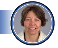
Angie Burns, MS, ACS, RCS
Assistant Professor / Clinical Coordinator
St. Catherine University
Angie Burns is an assistant professor and clinical coordinator in the sonography program at St. Catherine University (SCU) in St. Paul, MN. Before starting at SCU, Angie worked for 15 years as a cardiac sonographer at Mayo Clinical Health Systems, La Crosse, WI and as an advanced cardiac sonographer at Mayo Clinic, Rochester, MN. Angie earned her bachelor's degree and master's degree from the University of WI, La Crosse (UWL) where she studied clinical exercise physiology and cardiac rehabilitation. In addition to presenting at numerous local and national conferences, Angie has served in a volunteer capacity on the Events Management Committee (EMC) and as a CME reviewer for SDMS.
OB/GYN Track | 1.0 SDMS CME Credit | Category: OB | Content Level: Intermediate
Screening for aneuploidy is recommended for all Pregnancies regardless of maternal age or history. This presentation will discuss the role of ultrasound and the identification of soft markers and how those markers modify counseling to the patient and additional testing.
Objectives:
- Discuss the role of ultrasound in screening for aneuploidy.
- Describe the pathophysiology of soft markers and their functional significance.
- Clarify the role of soft markers of aneuploidy in the setting of universal aneuploidy screening.
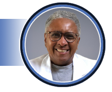
Lisa Moore, MD, RDMS
Professor
Texas Tech at Permian Basin
Lisa Moore, MD, RDMS is a board-certified perinatologist and sonographer with the Obstetric (OB) and Fetal Echo (FE) credentials. Dr. Moore attended medical school at Duke University followed by residency in Obstetrics and Gynecology at the Medical College of Georgia and fellowship in Maternal-Fetal Medicine at the University of Mississippi in Jackson. She has published in the Journal of Diagnostic Medical sonography (JDMS) and serves as a reviewer for the Journal. Dr. Moore is currently employed at Texas Tech Health Sciences center of the Permian Basin. She has experience in the interpretation of high-risk obstetrics ultrasound images and the management of ultrasound findings. She has extensive experience in organizing and managing an ultrasound unit.
Vascular Track | 1.0 SDMS CME Credit | Category: VT | Content Level: Intermediate
This presentation will bring awareness to Thoracic Outlet Syndrome and provide understanding to how to appropriately assess for the disease.
Objectives:
- Define and provide an overview of Thoracic Outlet Syndrome.
- Understand the causes, risk factors, and symptoms of TOS.
- Understand how to appropriately test for TOS and learn how to help these patients.

Aften Ferguson, MBA, RDMS, RDCS, RVT
Supervisor CV Testing & Diagnostic US
IU Health Bloomington
Aften Ferguson, MBA, RDMS, RDCS, RVT is the Supervisor of Cardiovascular Testing and Diagnostic Ultrasound at IU Health Bloomington located in Bloomington, IN. She's been with the organization for 10 years and in a supervisor role for three years. In addition, she is a strong supporter of numerous sonography programs in Indiana, is on the Advisory Board for a local community college in Indiana, and serves as a clinical instructor for several programs. Aften has a bachelor’s degree in Diagnostic Medical Sonography, and a master’s degree in healthcare management. She is very excited to start her journey working with the SDMS.
Abdominal + Track | 1.0 SDMS CME Credit | Category: AB | Content Level: Beginner
AFP, ALT, AST, GGT, TE, ARFI, LFT, PT, INR, MASH, MASLD - what are all these acronyms and how do they relate to the liver?
Objectives:
- Discuss the role of ultrasound in elevated liver function tests.
- Describe the different types of elastography related to the liver.
- Discuss the microscopic anatomy of the liver.

M. Robert DeJong, RDMS, RDCS, RVT, FAIUM, FSDMS
Owner
Bob DeJong, LLC.
Bob graduated from The Johns Hopkins School of Radiologic Technology in 1976. His career began with learning ultrasound, leading to over 25 years as the technical manager of the radiology ultrasound division at The Johns Hopkins Hospital. He now lectures at local and national ultrasound meetings and runs his own company, Bob DeJong, LLC, providing ultrasound education. He authored "Craig’s Essentials of Sonography and Patient Care" in 2018 and "Sonography Scanning: Principles and Protocols 5th Edition" in 2020, alongside contributing to numerous ultrasound textbooks. Bob received the Joan Baker Pioneer Award from the SDMS and delivered the Stephen McLaughlin Memorial Lecture at the 2022 SDMS Annual Conference. He holds fellow member status in both the SDMS and the AIUM, serving on the Board of Directors for the SDMS and SDMS Foundation. Married to Linda since 1975, they have two sons, Alex and Daniel, who have supported his career. In his free time, he is active in his church, enjoys theater, reading, board games, and is a Disney fan.
Cardiac Track | 1.0 SDMS CME Credit | Category: AE | Content Level: Intermediate
COVID-19 and long-term COVID-19 have led to various cardiac and other organ complications. This presentation offers an overview of COVID-19, its impact on the heart and other organs, and the use of electrocardiography (ECG) and echocardiography (ECHO) in diagnosing and managing pericardial diseases.
Objectives:
- Define COVID and how it affects the heart.
- Define Myocarditis and Long COVID.
- Review electrocardiogram (ECG) and echocardiogram (ECHO) findings.

Georgeanne Lammertin, MBA, RCS, RDCS, FASE
Sonographer
HCA
Georgeanne Lammertin is currently employed as a sonographer with HCA Florida. Her career spans over several prestigious institutions, beginning with 18.5 years at the University of Chicago, where she worked as a sonographer, researcher, and technical director. Following this, she spent 6 years at the University of Utah, serving as the manager of the Cardiovascular service line for both the University of Utah and Huntsman Cancer Center. Subsequently, Georgeanne transitioned to MedStar Washington, where she held the position of technical director of cardiovascular services for 2 years. Throughout her career, she has been actively involved in volunteer work, contributing her expertise to organizations such as the Society of Diagnostic Medical Sonography (SDMS), the American Society of Echocardiography (ASE), and Team Heart Rwanda.
OB/GYN Track | 1.0 SDMS CME Credit | Category: OB | Content Level: Intermediate
In this introspective and compassionate dialogue, Hayley calls for the ultrasound community to reflect upon how their personal biases create barriers to empathy and connection with patients, using real world examples. Using the World Health Organization supported principles of patient-centered care, Hayley examines how obstetrical sonographers play a critical role in health outcomes for vulnerable patients and discusses strategies for implementing supportive and sensitive care environments and advocacy for their patients.
Objectives:
- Acknowledge and appreciate the significance of the role sonographers play in the context of our patients' obstetric care.
- Discover ways to translate this powerful awareness into actionable strategies of patient-centered care practices in the ultrasound examination room.
- Understand how the patient experience directly impacts community health outcomes and the influential impact sonographer bias and communication can have in the obstetric setting.

Hayley Bartkus, MS-HPEd., BSDMS, RDMS
Diagnostic Medical Sonography Program Director
Johns Hopkins School of Medical Imaging
Hayley O. Bartkus is the Diagnostic Medical Sonography Program Director at the Johns Hopkins Schools of Medical Imaging. She graduated cum laude with a Bachelor of Science in Diagnostic Medical Sonography from the Rochester Institute of Technology in 2017, and later earned a Master of Science in Health Professions Education from Boston University Chobanian & Avedisian School of Medicine in 2022. With a passion for ultrasound education and patient-centered care, Hayley began her career at Albany Medical Center in 2017 before transitioning to the Hospital of the University of Pennsylvania, specializing in high-risk obstetric sonography. During her time at UPenn, she engaged in cutting-edge clinical research and played a key role in ultrasound education. Hayley is dedicated to advancing the field of diagnostic medical sonography, advocating for patient outcomes, and hosts the podcast "256 Shades of Gray". Recognized as a founding mentor at EchoMentor and honored with AS Software’s 2023 Sonography Impact Community Advocate Award, Hayley continues her mission of advocating for equitable, evidence-based medical care with a focus on sonographers and high-quality ultrasonic imaging.
Vascular Track | 1.0 SDMS CME Credit | Category: OT | Content Level: Intermediate
This presentation will cover climate change and its Impact on medical imaging.
Objectives:
- Explore the implications of climate change on epidemiology.
- Understand how these implications will affect healthcare workers.
- Understand these effects to the medical imaging technologist.

Paul English, BS, MS, RT(N), CNMT, RVT
Adjunct Professor
Dallas College
Paul English retired after 47 years in medical imaging, primarily in ultrasound and nuclear medicine. Initially considering a career as a research physicist, he became a mobile nuclear medicine technologist in 1974. Introduced to ultrasound in 1976, he later added CT to his repertoire. Throughout his career, Paul has been involved in teaching ultrasound and nuclear medicine students, second only to his passion for patient care. He has held various offices in national, state, and local ultrasound and nuclear medicine societies, and has given numerous presentations at various meetings and institutions. While he retired from clinical practice in 2020, he continues to teach physics for the Diagnostic Medical Sonography program at Dallas College as an adjunct instructor.
Abdominal + Track | 1.0 SDMS CME Credit | Category: PS | Content Level: Intermediate
This presentation will explore the pivotal role of ultrasound imaging in enhancing the assessment of pediatric abdominal trauma. The aim of this presentation is to offer a thorough comprehension of common causes of abdominal trauma in pediatric patients, as well as to delineate the typical clinical pathway utilized in the assessment. Additionally, the presentation will cover how ultrasound imaging, including the utilization of contrast-enhanced ultrasound imaging (CEUS), can assist in diagnosing injuries related to abdominal trauma in pediatric patients. Join to learn more about the complexities of assessing pediatric abdominal trauma and discover how ultrasound imaging improves diagnostic accuracy in these critical cases.
Objectives:
- Identify common causes of abdominal trauma within the pediatric population.
- Outline the common clinical pathway when assessing pediatric abdominal trauma.
- Discuss how ultrasound imaging aids in the diagnosis of common injuries associated with abdominal trauma pediatric patients, including the use of contrast enhanced ultrasound imaging (CEUS)

Cara Hill, BS, RT (R), RDMS, RVT
Clinical Education Specialist
GE HealthCare
With six years of hands-on experience in sonography, primarily focusing in pediatrics at Children’s National Hospital in Washington, DC, and four years as an educator in Diagnostic Medical Sonography programs at institutions such as Northern Virginia Community College and her alma mater, Misericordia University, Cara Hill brings expertise and passion to the forefront of diagnostic ultrasound. Her commitment extends beyond the clinic and classroom, as she actively volunteers her time and expertise in global health initiatives with RAD-AID International. In 2021, Cara took on the role of Program Manager for the RAD-AID Laos Project after quickly learning that volunteering with RAD-AID allows the demonstration of several important values, including commitment, engagement, compassion, empathy, and respect for others. As a dedicated member of the Society of Diagnostic Medical Sonography (SDMS), Cara actively contributes to the advancement of the field and stays abreast of the latest developments and best practices. She continuously seeks opportunities to mentor and inspire the next generation of sonographers.
Cardiac Track | 1.0 SDMS CME Credit | Category: AE | Content Level: Intermediate
Echocardiography is an invaluable tool for the assessment of cardiac masses. Masses found within the heart can include thrombus, vegetations, benign or malignant tumors, normal variants, or extra cardiac structures. In this presentation, we will describe various tumors and masses found in the heart and discuss some of the echocardiographic features that accompany them.
Objectives:
- Describe the echocardiographic characteristics of an intra cardiac thrombus.
- Identify the most common locations of cardiac tumors.
- Describe the echocardiographic features that help to differentiate benign versus malignant tumors.

Angie Burns, MS, ACS, RCS
Assistant Professor / Clinical Coordinator
St. Catherine University
Angie Burns is an assistant professor and clinical coordinator in the sonography program at St. Catherine University (SCU) in St. Paul, MN. Before starting at SCU, Angie worked for 15 years as a cardiac sonographer at Mayo Clinical Health Systems, La Crosse, WI and as an advanced cardiac sonographer at Mayo Clinic, Rochester, MN. Angie earned her bachelor's degree and master's degree from the University of WI, La Crosse (UWL) where she studied clinical exercise physiology and cardiac rehabilitation. In addition to presenting at numerous local and national conferences, Angie has served in a volunteer capacity on the Events Management Committee (EMC) and as a CME reviewer for SDMS.
OB/GYN Track | 1.0 SDMS CME Credit | Category: OB | Content Level: Advanced
This presentation will cover all aspects of creating and managing an obstetric ultrasound unit for the private practice office including quality control, imaging protocols, scheduling, staffing, image storage, physician interpretation, result reporting, quality metrics and patient satisfaction.
Objectives:
- Describe the basic requirements for a functional obstetric ultrasound unit.
- Describe basic principles of protocol-based image acquisition, storage and interpretation.
- Describe the functional role of physicians and sonographers in the obstetric ultrasound unit.

Lisa Moore, MD, RDMS
Professor
Texas Tech at Permian Basin
Lisa Moore, MD, RDMS is a board-certified perinatologist and sonographer with the Obstetric (OB) and Fetal Echo (FE) credentials. Dr. Moore attended medical school at Duke University followed by residency in Obstetrics and Gynecology at the Medical College of Georgia and fellowship in Maternal-Fetal Medicine at the University of Mississippi in Jackson. She has published in the Journal of Diagnostic Medical sonography (JDMS) and serves as a reviewer for the Journal. Dr. Moore is currently employed at Texas Tech Health Sciences center of the Permian Basin. She has experience in the interpretation of high-risk obstetrics ultrasound images and the management of ultrasound findings. She has extensive experience in organizing and managing an ultrasound unit.
Vascular Track | 1.0 SDMS CME Credit | Category: OT | Content Level: Intermediate
This presentation will define work-related musculoskeletal injuries, how to prevent them, and how to put ergonomics first.
Objectives:
- Define and explain musculoskeletal injuries.
- Learn that prevention is the key! Stretch happens!
- Put ergonomics first and prevent injury for yourself.

Aften Ferguson, MBA, RDMS, RDCS, RVT
Supervisor CV Testing & Diagnostic US
IU Health Bloomington
Aften Ferguson, MBA, RDMS, RDCS, RVT is the Supervisor of Cardiovascular Testing and Diagnostic Ultrasound at IU Health Bloomington located in Bloomington, IN. She's been with the organization for 10 years and in a supervisor role for three years. In addition, she is a strong supporter of numerous sonography programs in Indiana, is on the Advisory Board for a local community college in Indiana, and serves as a clinical instructor for several programs. Aften has a bachelor’s degree in Diagnostic Medical Sonography, and a master’s degree in healthcare management. She is very excited to start her journey working with the SDMS.